Ravi

mri-knee-meniscus-degeneration-maceration-flap-tear-extrusion-meniscocapsular-separation-radedasia

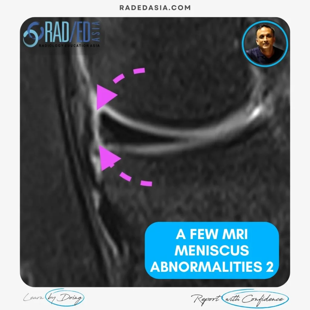
A FEW MRI MENISCUS ABNORMALITIES 2: MRI MENISCUS DEGENERATION MACERATION FLAP TEAR EXTRUSION AND MENISCOCAPSULAR SEPARATION

MENISCUS ABNORMALITIES 2 KNEE MRI MRI MENISCUS ABNORMALITIES A few more MRI Meniscus abnormalities such as meniscus maceration, flap tears, extrusion and meniscocapsular separation. MRI MENISCUS ABNORMALITIES MENISCO CAPSULAR SEPARATION MRI Meniscocapsular separation is a separation of the attachment of the external margin of the body of the medial meniscus from the Posterior Oblique ligament. […]

A FEW MRI MENISCUS ABNORMALITIES 2: MRI MENISCUS DEGENERATION MACERATION FLAP TEAR EXTRUSION AND MENISCOCAPSULAR SEPARATION Read More »

ankylosing spondylitis spine ct radiology

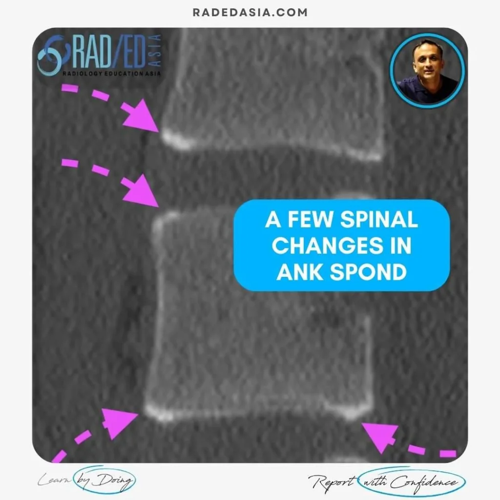
A FEW SPINAL CHANGES IN ANKYLOSING SPONDYLITIS

ANKYLOSING SPONDYLITIS SPINAL CHANGES CT & MRI Findings in Ankylosing Spondylitis of the spine Ankylosing spondylitis (AS), in the spine can involve the anterior and posterior elements. It usually commences anteriorly and as the disease progresses, posterior changes are seen. In these images we look at: Corner erosions and Sclerosis.  Syndesmophyte formation between vertebrae. Fusion:

A FEW SPINAL CHANGES IN ANKYLOSING SPONDYLITIS Read More »

mri knee meniscus tears cysts pathologies images

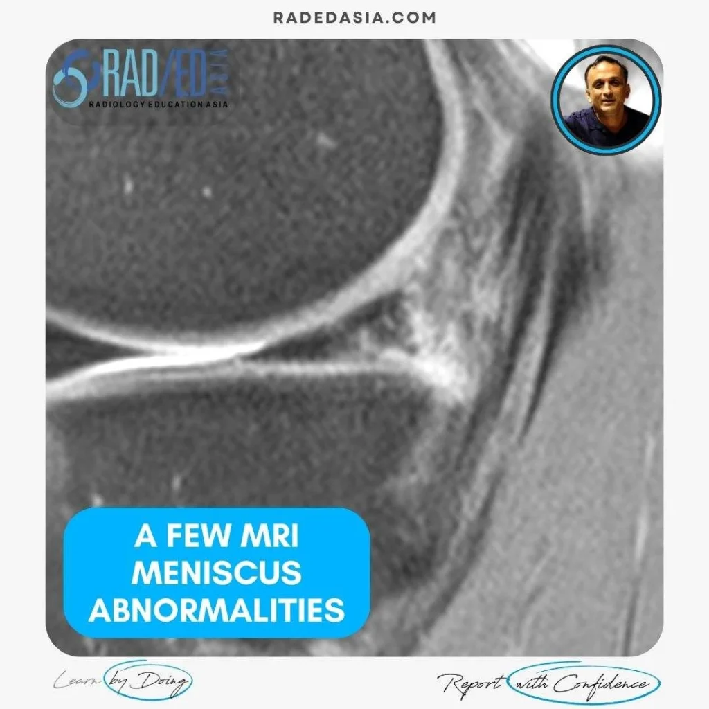
A FEW MRI MENISCUS ABNORMALITIES: MENISCUS TEAR, INTRA MENISCAL CYST AND VARIANTS THAT LOOK LIKE TEARS

MENISCUS ABNORMALITIES KNEE MRI MRI MENISCUS ABNORMALITIES A few MRI Meniscus abnormalities such as Meniscus tear, Intra meniscal cyst and variants that look like tears. MRI MENISCUS ABNORMALITIES MRI MENISCUS INTRAMENISCAL CYST WHAT ARE THE FINDINGS? Cystic structure (Pink arrow) present within the anterior horn. WHAT’S THE Dx? INTRAMENISCAL CYST Extensive intra meniscal cyst anterior

A FEW MRI MENISCUS ABNORMALITIES: MENISCUS TEAR, INTRA MENISCAL CYST AND VARIANTS THAT LOOK LIKE TEARS Read More »

mri achilles tendon abnormalities ankle radiology

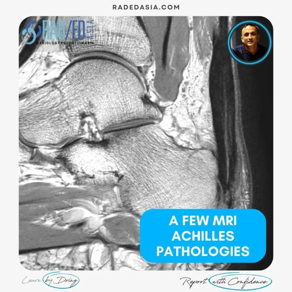
MRI OF A FEW ACHILLES TENDON ABNORMALITIES: INSERTIONAL TENDONOPATHY TENDONOSIS, BURSA & RUPTURE

ACHILLES TENDON ABNORMALITIES ANKLE MRI ACHILLES TENDON ABNORMALITIES A few MRI images of Achilles pathology from all the findings seen in Achilles Insertional Tendonopathy, Retrocalcaneal Bursitis, Retroachilles Bursitis to MRI of Achilles Rupture. MRI ACHILLES TENDON ABNORMALITIES MRI ACHILLES INSERTIONAL TENDONOPATHY: WHAT TO LOOK FOR MRI findings in Insertional Achilles Tendinopathy: A few findings to

MRI OF A FEW ACHILLES TENDON ABNORMALITIES: INSERTIONAL TENDONOPATHY TENDONOSIS, BURSA & RUPTURE Read More »

polyarteritis nodosa myopericarditis myositis mri pet ct coronary angiogram imaging radedasia

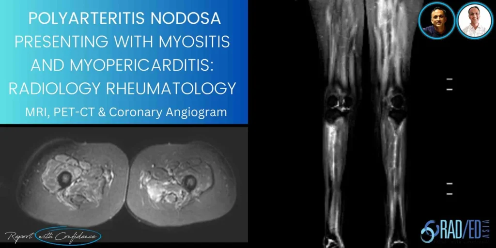
POLYARTERITIS NODOSA WITH MYOPERICARDITIS AND MYOSITIS : MRI PET CT & CORNARY ANGIOGRAM FINDINGS

INTRODUCTION A combined Clinico Radiological case from Dr Joe Thomas (Rheumatologist) This is a combined case with Dr Joe Thomas on a case of Polyarteritis Nodosa that presented with Myositis and Myopericarditis. Dr Thomas is a senior consultant Rheumatologist who is also joining us in presenting the Spine Arthropathy and Spondyloarthropathy course. PATIENT HISTORY FIRST

POLYARTERITIS NODOSA WITH MYOPERICARDITIS AND MYOSITIS : MRI PET CT & CORNARY ANGIOGRAM FINDINGS Read More »

pyriformis syndrome pudendal nerve quadratus femoris ischiofemoral impingement ultrasound scan inject radedasia

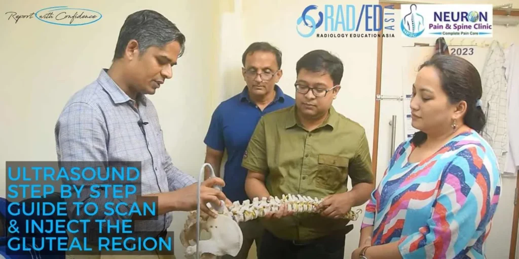
ULTRASOUND PAIN MANAGEMENT HOW TO SCAN AND INJECT FOR PIRIFORMIS SYNDROME, PUDENDAL NERVE, QUADRATUS FEMORIS & ISCHIOFEMORAL IMPINGEMENT

STEP BY STEP GUIDE ON HOW TO SCAN AND INJECT THE GLUTEAL REGION FOR PAIN MANAGEMENT HOW TO SCAN AND INJECT THE GLUTEAL REGION FOR PAIN MANAGEMENT   In this video by Dr Rajendra Sahoo Pain Management Specialist at KIMS Hospital and Neuron Pain Clinic we look at the role of ultrasound in the diagnosis

ULTRASOUND PAIN MANAGEMENT HOW TO SCAN AND INJECT FOR PIRIFORMIS SYNDROME, PUDENDAL NERVE, QUADRATUS FEMORIS & ISCHIOFEMORAL IMPINGEMENT Read More »

philips learning academy APAC radedasia

PHILIPS APAC LEARNING ACADEMY COLLABORATION MSK SPINE MRI COURSE

RADEDASIA & PHILIPS APAC LEARNING ACADEMY: A NEW COLLABORATION ALL 8 OF OUR ONLINE GUIDED MRI MINI FELLOWSHIPS NOW ON THE PHILIPS APAC LEARNING ACADEMY We are commencing a new collaboration with Philips for all our Guided Online MRI Mini Fellowships to be on the Philips APAC Learning Academy. If you are a Philips Customer

PHILIPS APAC LEARNING ACADEMY COLLABORATION MSK SPINE MRI COURSE Read More »

meniscal ossicles mri knee radiology education asia

MENISCAL OSSICLES: POSTERIOR HORN MENISCUS PART 3

MENISCAL OSSICLES: POSTERIOR HORN MENISCUS MENISCAL OSSICLES: WHAT ARE THEY Meniscal ossicles are areas of ossification that occur within the meniscus. WHY DO THEY OCCCUR Two possible causes are given: They are a Result of Post traumatic metaplasia and heterotopic ossification of a meniscal tear (either posterior horn or root). This is the most likely

MENISCAL OSSICLES: POSTERIOR HORN MENISCUS PART 3 Read More »

acromial spur mri shoulder radiology education asia

ACROMION / ACROMIAL SPURS ON MRI: WHERE TO LOOK AND WHAT TO LOOK FOR

ACROMION SPURS ON MRI: 3 APPEARANCES ON MRI   Acromial / Acromion spurs can be quite small and difficult to visualize on MRI.   They are enthesophytes that arise from traction at the attachment sites of the Coraco Acromial Ligament and Deltoid.   This post looks at the three locations and most common appearances of

ACROMION / ACROMIAL SPURS ON MRI: WHERE TO LOOK AND WHAT TO LOOK FOR Read More »

error: Content is protected !!

Join our Newsletter

Stay tuned on new
Mini-Fellowships launches and learnings

Mini-Fellowships

Better Than Conferences

Join our WhatsApp Community

We help radiologists learn
by doing and report with confidence

This site is intended for Medical Professions only. Use of this site is governed by our Terms of Service and Privacy Statement which can be found by clicking on the links. Please accept before proceeding to the website.