How to diagnose uncovertebral joint (uncinate process) degeneration on cervical spine CT — coronal findings, sclerosis, and foraminal nerve root compression.

Stay tuned on new Fellowships and learnings

CERVICAL CT: UNCOVERTEBRAL JOINT DEGENERATION — HOW TO DIAGNOSE

CERVICAL CT: UNCOVERTEBRAL JOINT DEGENERATION

How do you diagnose uncovertebral joint degeneration on CT?

  • The best way to see uncovertebral joint degeneration is on CT, starting with the normal appearance of the joint before looking for degenerative change.
  • The coronal CT is the best plane to assess for uncovertebral degeneration.
  • In this video we go through normal uncovertebral joint anatomy, the CT appearance of degeneration, and why identifying it matters clinically.

If you find the video helpful, please subscribe to the channel.

NORMAL UNCOVERTEBRAL JOINT ANATOMY
  • The uncinate process arises from the vertebral body and articulates with the inferolateral aspect of the vertebral body above.
  • The inferior margin of the vertebral body above has a beveled or angled appearance — generally referred to as the articular margin of the uncovertebral joint.
  • There has been debate over whether this is a true joint, but it is now considered a proper joint, with some fibrocartilage present.
  • It is a synovial joint, so it is prone to inflammatory change.
  • Its main purpose is to restrict lateral flexion of the neck.
  • The joint forms the anterior margin of the intervertebral foramen, which is why degeneration here has downstream effects on the exiting nerve root.
HOW TO IDENTIFY DEGENERATION ON CT

On the normal side, there is a nice, well-preserved space between the uncinate process and the inferior articular margin, with no degenerative change.

  • Narrowing of the joint space.
  • Significant sclerosis at either side of the articular margin.
  • Hypertrophic change involving the whole region, including the uncinate process, best appreciated by comparison with the contralateral, normal side.

Coronal reformats are used first to identify the degeneration itself, and sagittal reformats are then used to assess for foraminal narrowing from osteophyte formation arising from the degenerated joint.

WHY IT MATTERS
  • Osteophytes arising from uncovertebral joint degeneration extend into the intervertebral foramen and can compress the exiting nerve root.
  • The degenerated joint can also be a pain generator in its own right, independent of any nerve root compression.
KEY POINT
  • Uncovertebral joint degeneration is best seen on CT, predominantly on coronal reformats.
  • Degenerative findings include joint space narrowing, sclerosis at the articular margin, and hypertrophic change of the uncinate process, best appreciated against the contralateral normal side.
  • Osteophytes from this joint extend into the intervertebral foramen and can compress the exiting nerve root — check sagittal reformats for foraminal narrowing.
  • The uncovertebral joint itself can also be a pain generator, independent of any nerve root compression.
TEST YOURSELF ON SOME COMMON & FREQUENTLY ASKED QUESTIONS
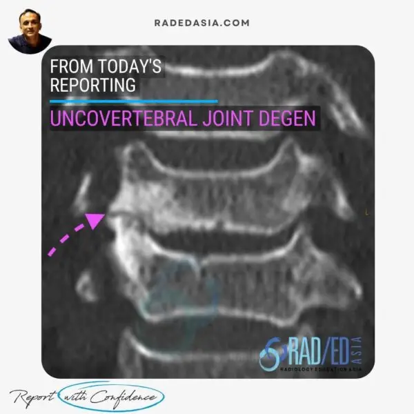
Coronal CT reformats. Start there to identify the degeneration, then check sagittal reformats for any resulting foraminal narrowing.

Narrowing of the joint space, significant sclerosis at either side of the articular margin, and hypertrophic change of the uncinate process, compared against the normal contralateral side.

Two reasons: osteophytes arising from the degenerated joint can extend into the intervertebral foramen and compress the exiting nerve root, and the degenerated joint itself can be a pain generator on its own.

If you wish to watch the video in language other than English, please view it on our YouTube Channel HERE

We look at all of these topics in more detail in our Guided SPINE & SIJ Imaging Mini Fellowships.

Click on the image below for more information.

Our CPD & Learning Partners

OTHER POPULAR SPINE REPORTING POSTS: CLICK ON THE IMAGES BELOW

CT scan of the cervical spine showing uncovertebral joint degeneration with sclerosis and loss of joint space coronal reformat
PAIN MANAGEMENT
annular fissure tear mri radiology lumbar spine radedasia
Spanish Blogs

Stay tuned on new Fellowships and learnings

error: Content is protected !!

Join our Newsletter

Stay tuned on new
Mini-Fellowships launches and learnings

Mini-Fellowships

Better Than Conferences

Join our WhatsApp Community

We help radiologists learn
by doing and report with confidence

This site is intended for Medical Professions only. Use of this site is governed by our Terms of Service and Privacy Statement which can be found by clicking on the links. Please accept before proceeding to the website.