chondromalacia patella femoral trochlear knee cartilage mri radiology radedasia

CHONDROMALACIA PATELLA FEMORAL TROCHLEAR CARTILAGE MRI KNEE

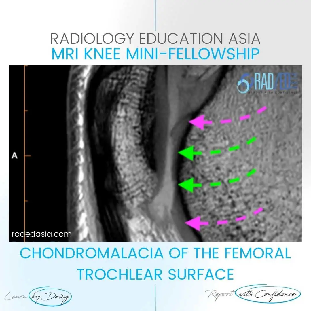
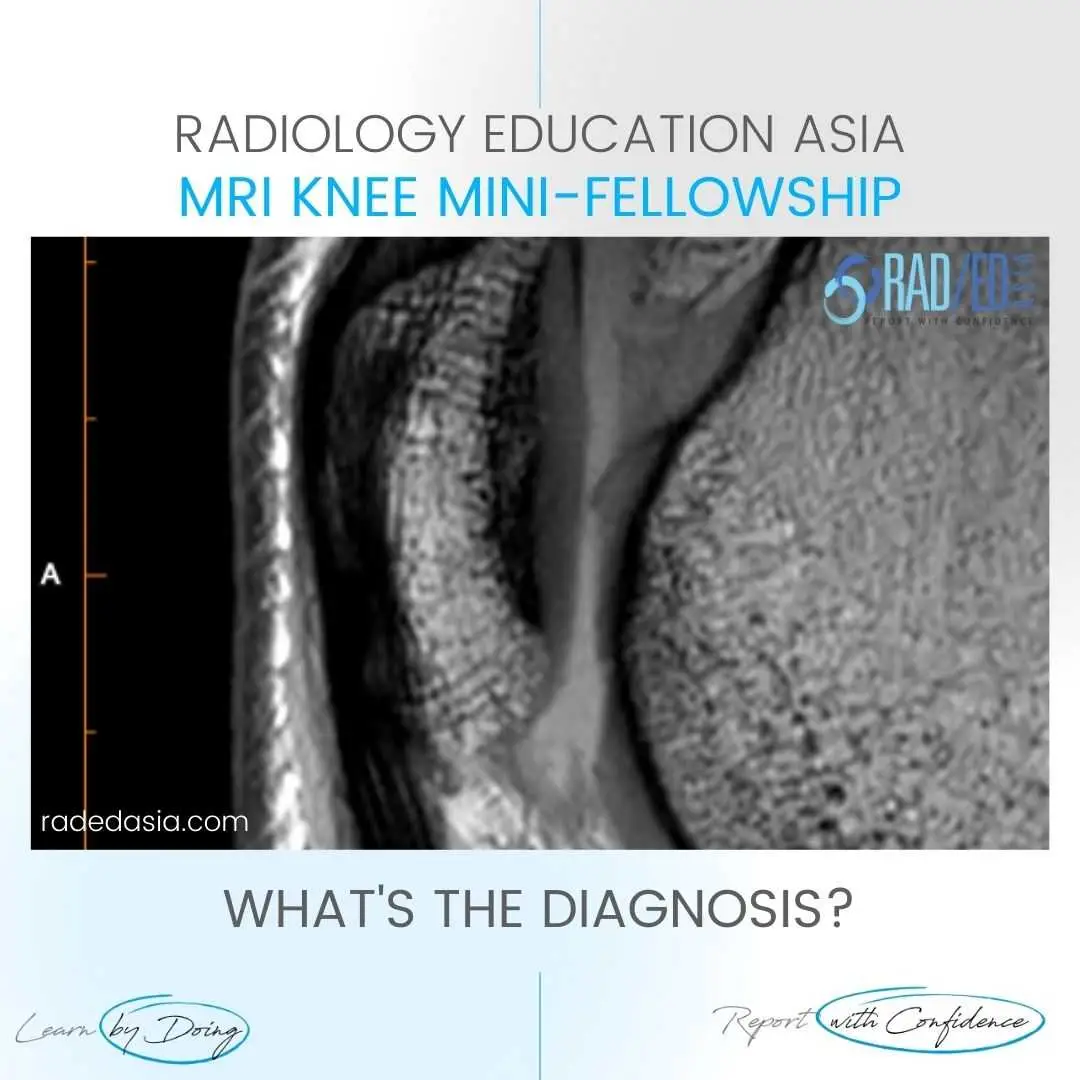
  • Chondromalacia of the femoral trochlear surface.


  • There is severe (in areas full thickness) cartilage loss (Green arrows) in the femoral trochlear.


  • Compare with normal thickness cartilage above and below (Pink arrows).


  • The femoral trochlear surface cartilage should be assessed on axial and Sagittal scans.

Severe cartilage loss (Green arrows).  Compare with normal thickness cartilage (Pink arrows).
chondromalacia-patella-femoral-trochlear-knee-cartilage-mri-radiology-radedasia.jpgchondromalacia-patella-femoral-trochlear-knee-cartilage-mri-radiology.jpg

#radiology #radedasia #mri #kneemri #msk #mriknee #mskmri #radiologyeducation #radiologycases #radiologist #rads #radiologystudent #radiologycme #radiologycpd #medicalimaging #imaging #radcme #rheumatology #arthritis #rheumatologist #sportsmed #orthopaedic #physio #physiotherapist #chondromalaciapatella

error: Content is protected !!

Join our Newsletter

Stay tuned on new
Mini-Fellowships launches and learnings

Mini-Fellowships

Better Than Conferences

Join our WhatsApp Community

We help radiologists learn
by doing and report with confidence

This site is intended for Medical Professions only. Use of this site is governed by our Terms of Service and Privacy Statement which can be found by clicking on the links. Please accept before proceeding to the website.