de quervain's tenosynovitis wrist ultrasound how to scan radiology radedasia

DE QUERVAIN’S TENOSYNOVITIS: HOW TO SCAN ON ULTRASOUND (VIDEO)

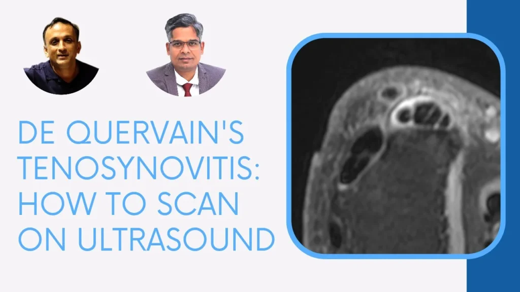
DE QUERVAIN’S TENOSYNOVITIS: HOW TO SCAN AND INJECT ON ULTRASOUND

  • De Quervain’s tenosynovitis is a stenosing tenosynovitis involving theĀ  retinaculum of the first dorsal compartment of the wrist, with involvement of abductor pollicis longus (APL) and extensor pollicis brevis (EPB) tendons and their synovial sheath.
  • Ultrasound assessment requires being able to identify the first dorsal compartment and its components.
  • In this video, Dr Rajendra Sahoo demonstrates where to look, what to look for and how to position the probe in assessing the first dorsal compartment of the wrist for De Quervain’s tenosynovitis on ultrasound.
  • Upcoming post will look at the MRI appearance of De Quervain’s tenosynovitis.

If you find the video helpful, please subscribe to the channel.

If your Browser is blocking the video, Please Click HERE to view it on our YouTube channel.

rajendra sahoo white bg pain management specialist radedasia 300px

Dr RAJENDRA SAHOO

Dr Rajendra Sahoo is a senior consultant Pain Physician and Anesthetist who is Fellowship trained in Ultrasound guided Pain Management. We will be collaborating with Dr Sahoo to bring more MSK and Spine Ultrasound learning.

Learn more about this condition & How best to report it in more detail in our

COMING SOON
Online Guided WRIST & HAND MRI Mini Fellowship

  • Join our WhatsApp Group for regular educational posts. Message “JOIN GROUP” to +6594882623 (your name and number remain private and cannot be seen by others).

  • Get our weekly email with all our educational posts: https://bit.ly/whathappendthisweek

#radedasia #mri #mskmri #radiology

This site is intended for Medical Professionals only. Use of this site is governed by our Terms of Service and Privacy Statement which can be found by clicking on the links. Please accept before proceeding to the website.
error: Right Click not available.