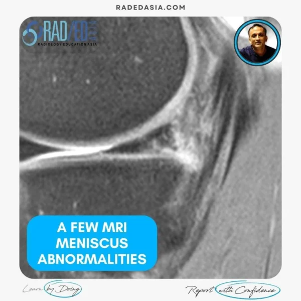
MRI MENISCUS ABNORMALITIES
A few more MRI Meniscus abnormalities such as meniscus maceration, flap tears, extrusion and meniscocapsular separation.![]()
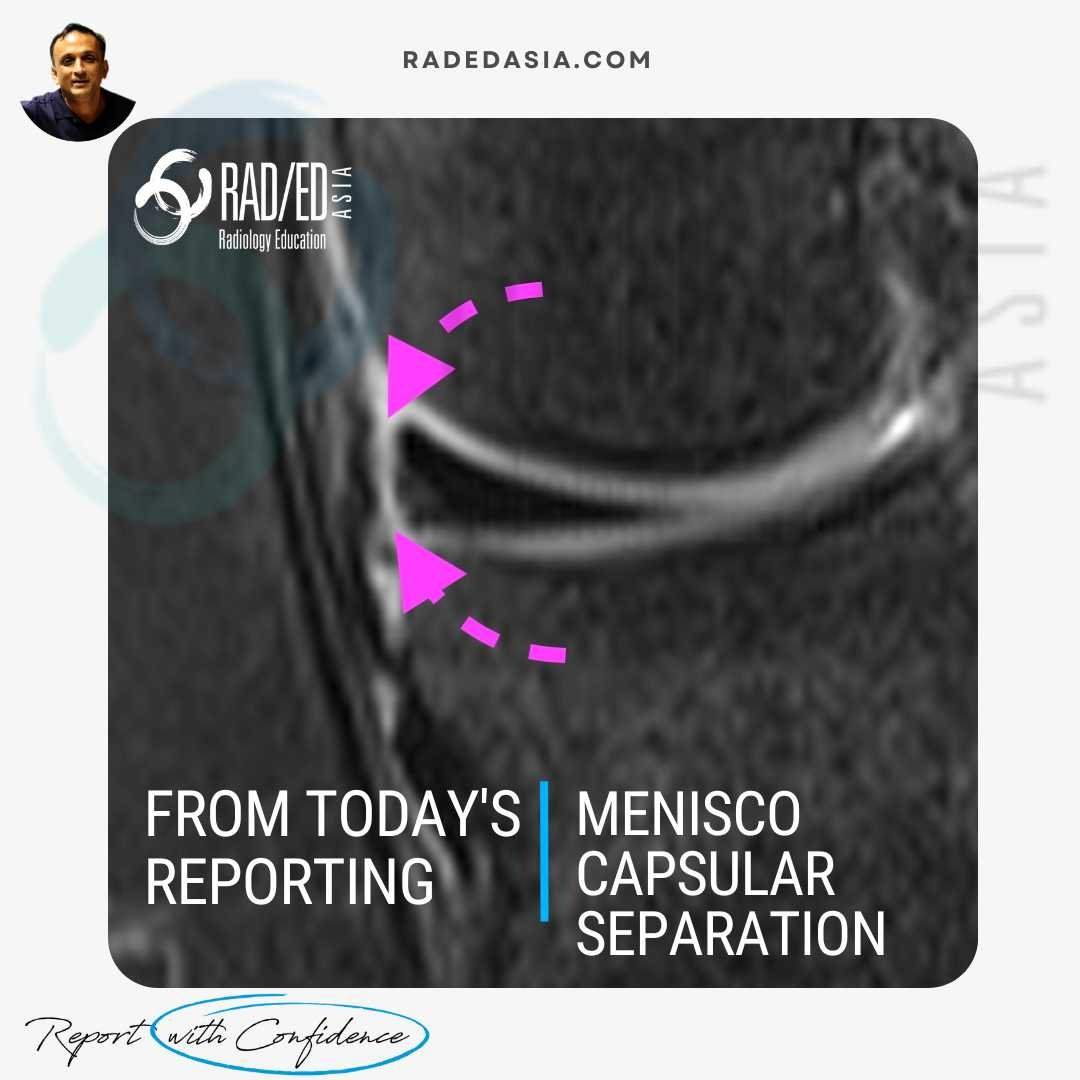
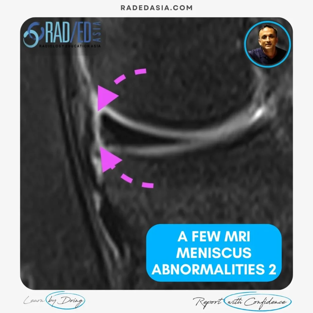
- Meniscocapsular separation is a separation of the attachment of the external margin of the body of the medial meniscus from the Posterior Oblique ligament.
Normally they are tightly attached and there should be no high signal between them.
Here we see a rim of high signal where the two have separated.
![]()

- A flap tear of the meniscus is a horizontal type tear where one side of the meniscus rotates away.
- Most commonly its in the medial meniscus body.
- One fragment is displaced out of the joint to lie between the sMCL and tibia or femur.
![]()

- Meniscal extrusion is when part of the meniscus is displaced from the joint.
- This is measured on a coronal scan.
- Displacement of the meniscus more than 2.5 - 3mm from the edge of the medial tibial plateau indicates extrusion.
- The two causes of extrusion are:
- Reduction in joint space from loss of cartilage.
- Tear of the posterior horn root.
Edge > 2.5 - 3.0 mm from Medial Tibial Plateau Margin).
![]()

- Maceration of the meniscus is the most severe form of meniscal degeneration. What to look for? Technical term alert…Looks like mush 😀
- Think of grinding up something in a mortar and pestle and that’s what you get in maceration.
- The meniscus generally retains it's shape but is ill defined and intermediate in signal.
- Due to the degeneration it's more likely to tear and it also stops functioning like a normal meniscus.
![]()