PAIN MANAGEMENT

Pain management Physician Anesthetist

All our posts on pain management are done in conjunction with Dr Rajendra Sahoo is a senior consultant Pain Physician and Anesthetist who is Fellowship trained in Ultrasound guided Pain Management & has a vast amount of clinical experience.

We will be collaborating with Dr Sahoo to bring more MSK and Spine Ultrasound learning.

subacromial subdeltoid bursa bursitis msk mri radiology radedasia

SUBACROMIAL SUBDELTOID BURSITIS MRI RADIOLOGY (VIDEO)

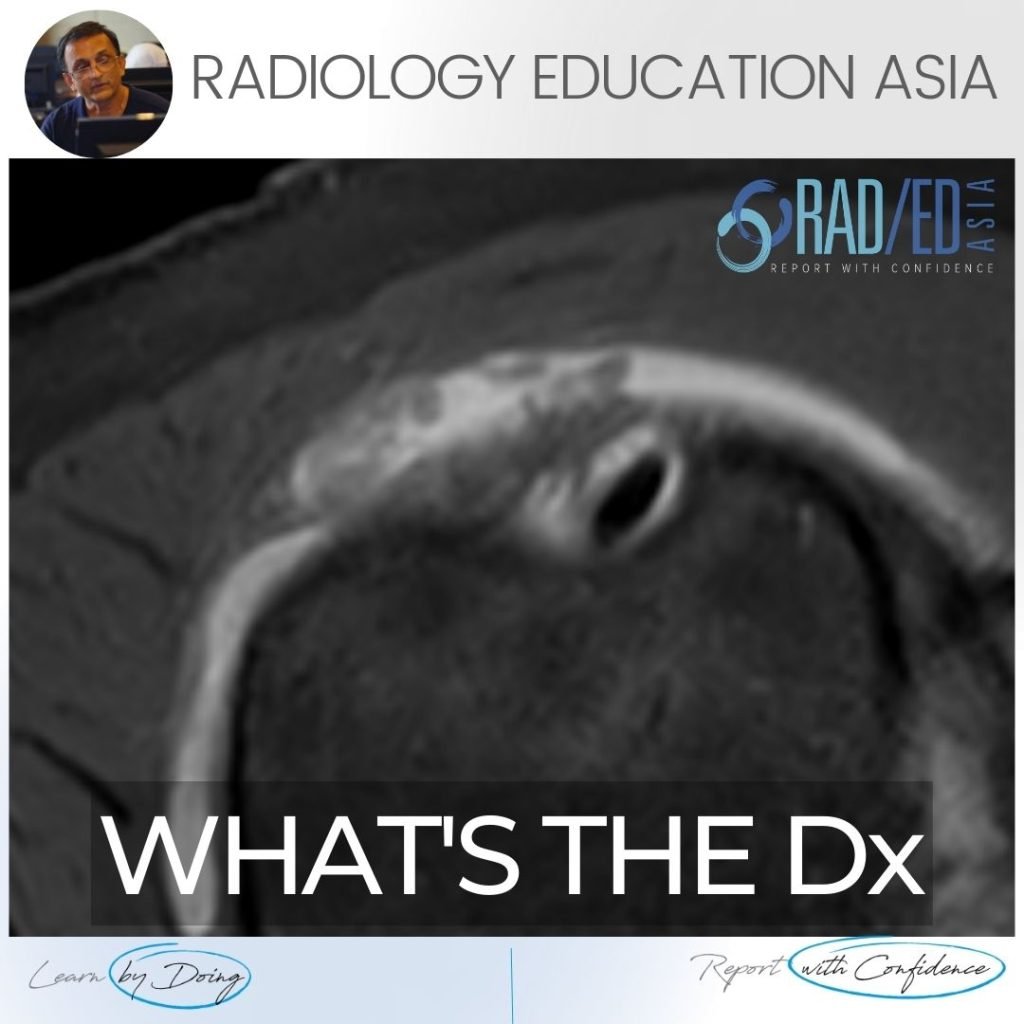
SUBACROMIAL SUBDELTOID BURSITIS MRI RADIOLOGY DISCUSSION Subacromial subdeltoid (SASD) bursitis on MRI can have a number of appearances. In this case we have a nodular type of synovitis which should raise the possibility of an inflammatory arthritis as a cause (Rheumatoid Arthritis in this case). WHAT DO WE SEE ON THE IMAGE? Fluid in the …

SUBACROMIAL SUBDELTOID BURSITIS MRI RADIOLOGY (VIDEO) Leer más »

mri ct osteitis condensans ilii spine sij

OSTEITIS CONDENSANS ILII RADIOLOGY CT SACROILIAC JOINT SIJ (VIDEO)

OSTEITIS CONDENSANS ILII RADIOLOGY CT SACROILIAC JOINT SIJ DISCUSSION ON OCI Osteitis Condensans Ilii.   Typical CT sclerotic changes of Osteitis Condensans Ilii. The sclerosis is continuous with the SIJ margin and particularly on the patient’s right it has a triangular appearance.   There is also mild sclerosis in the right sacrum adjacent to the …

OSTEITIS CONDENSANS ILII RADIOLOGY CT SACROILIAC JOINT SIJ (VIDEO) Leer más »

iliotibial band mri anatomy syndrome trauma mri knee radedasia

ILIOTIBIAL BAND MRI ANATOMY, SYNDROME, TRAUMA KNEE RADIOLOGY

ILIOTIBIAL BAND MRI ANATOMY, SYNDROME, TRAUMA Finding and assessing the Iliotibial Band (ITB) on MRI of the Knee is the first structure we assess when looking at the lateral side of the Knee.  In these three posts we look at: The normal MRI anatomy of the ITB. MRI of Iliotibial Band Syndrome (ITB Friction Syndrome) …

ILIOTIBIAL BAND MRI ANATOMY, SYNDROME, TRAUMA KNEE RADIOLOGY Leer más »

mri musculotendinous junction cysts and delaminating rotator cuff tears

MRI DELAMINATION ROTATOR CUFF TEARS MUSCULOTENDINOUS JUNCTION CYSTS

MRI DELAMINATING ROTATOR CUFF TEARS & MUSCULOTENDINOUS JUNCTION CYSTS On MRI Rotator cuff delaminating tears can result in Musculotendinous Junction Cysts.   These Musculotendinous junction cysts are seen in the rotator cuff tendons, mostly in Supraspinatus and less commonly in infraspinatus.   In this post we look at the appearance of both Delaminating Rotator cuff …

MRI DELAMINATION ROTATOR CUFF TEARS MUSCULOTENDINOUS JUNCTION CYSTS Leer más »

tibialis posterior tendinitis tendinopathy tear mri radiology

TIBIALIS POSTERIOR TENDINITIS TENDINOPATHY TEARS RADIOLOGY MRI (VIDEO)

TIBIALIS POSTERIOR TENDINITIS TENDINOPATHY TEARS RADIOLOGY MRI WHAT ARE THE FINDINGS The tibialis posterior tendon is enlarged and increased in signal. DIAGNOSIS & DISCUSSION TIBIALIS POSTERIOR TENDINOPATHY AND PARTIAL TEAR. Tibialis posterior tendinopathy and delamination / partial tears. The normal tibialis posterior tendon should be uniformly low signal. Enlargement and or increased intermediate signal indicates …

TIBIALIS POSTERIOR TENDINITIS TENDINOPATHY TEARS RADIOLOGY MRI (VIDEO) Leer más »

ischiofemoral impingement radiology mri hip

ISCHIOFEMORAL IMPINGEMENT RADIOLOGY HIP MRI (VIDEO)

ISCHIOFEMORAL IMPINGEMENT HIP MRI RADIOLOGY ISCHIOFEMORAL IMPINGEMENT: WHY DOES IT OCCUR Ischiofemoral impingement is a result of the quadratus femoris muscle being compressed between the ischial tuberosity and the posterior femur resulting initially in quadratus femoris oedema but can progress to tears and severe atrophy. ISCHIOFEMORAL IMPINGEMENT: WHAT ARE THE FINDINGS Initially there is oedema …

ISCHIOFEMORAL IMPINGEMENT RADIOLOGY HIP MRI (VIDEO) Leer más »

Este sitio está destinado únicamente a profesionales médicos. El uso de este sitio se rige por nuestra Términos de servicio y Declaracion de privacidad que se puede encontrar haciendo clic en los enlaces. Acepte antes de continuar con el sitio web.
error: Clic derecho no disponible.