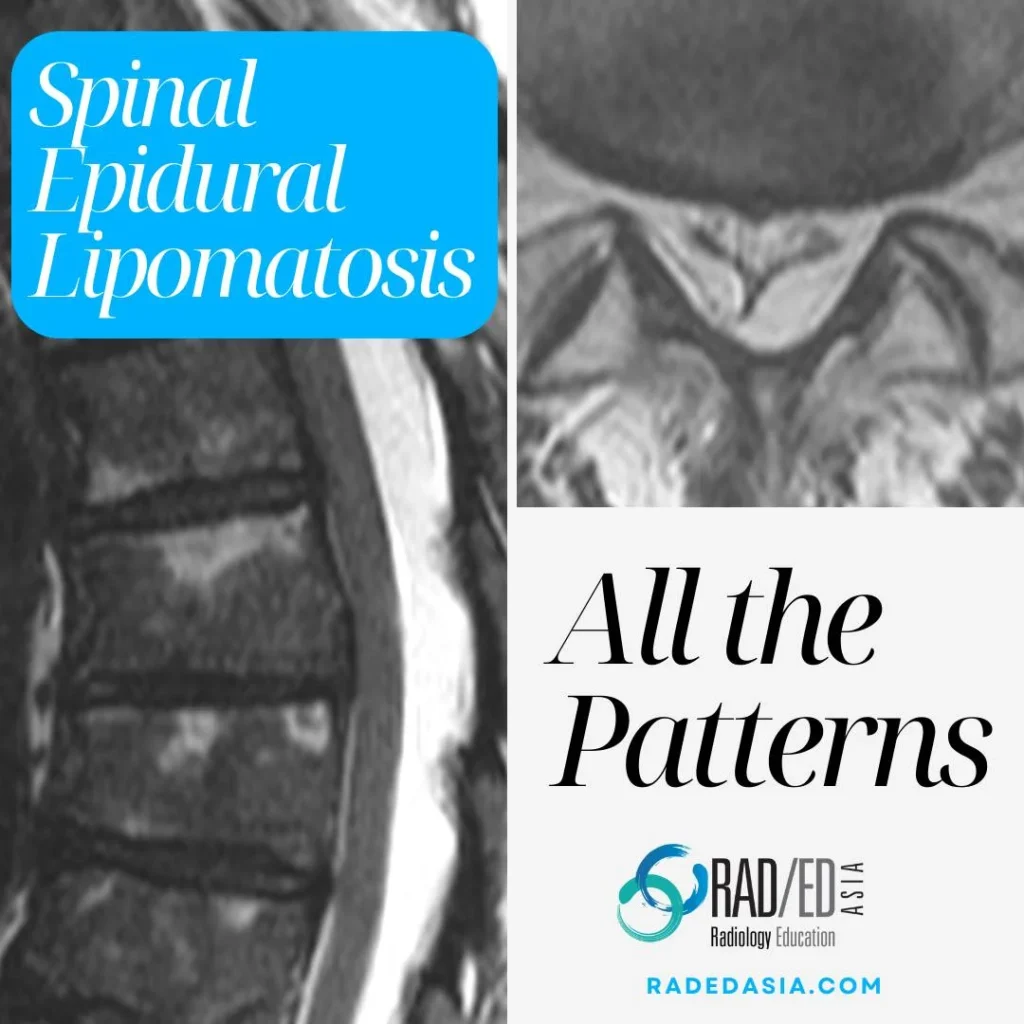
SPINAL EPIDURAL LIPOMATOSIS
- Spinal epidural lipomatosis is an excessive accumulation of non-encapsulated fat in the epidural space.
- It can be seen on MRI in the thoracic or lumbar spine. In the cervical spine epidural lipomatosis is not seen.
- Epidural lipomatosis is not uncommon to see and is an important diagnosis to make as it can initially be symptomatic.
- In this post we look at what are the characteristic appearances of lumbar and thoracic spinal epidural lipomatosis and how to make the diagnosis.
![]()
Often it's idiopathic but specific risk factors for developing Spinal Epidural lipomatosis are:
- Excessive exogenous or endogenous steroids.
- Spinal surgery.
- Obesity.

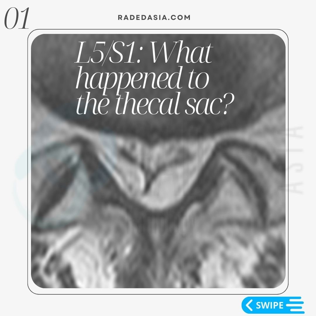
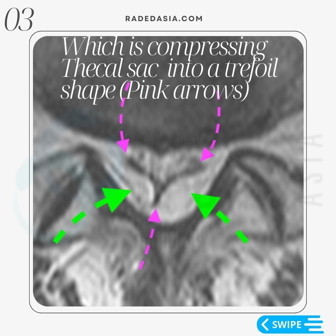
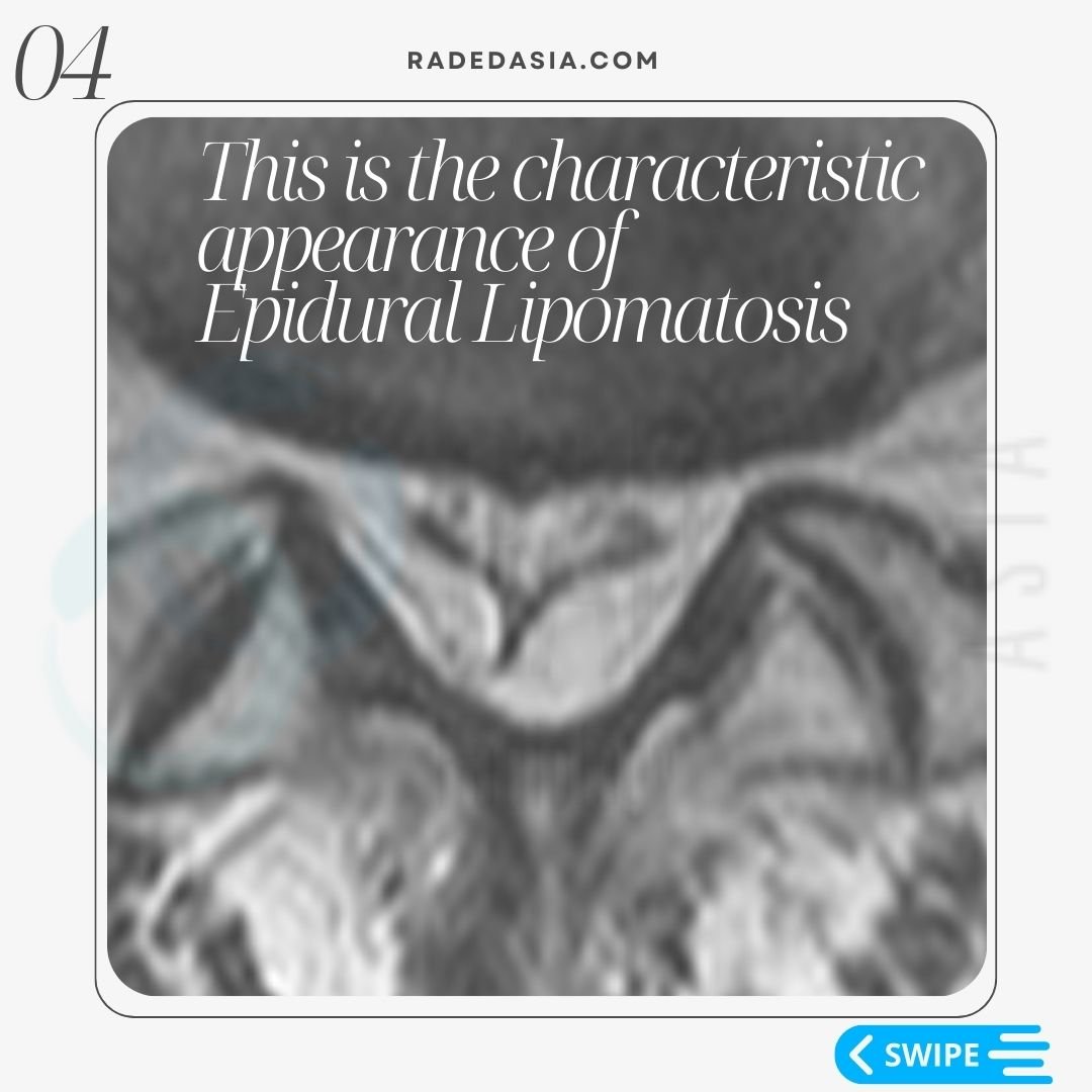
- The characteristic MRI finding in lumbar spinal epidural lipomatosis is a marked increase in fat in the epidural space (Green arrows in image below).
- This results in the thecal sac being compressed into a characteristic trefoil shape (Pink arrow image below).
- The trefoil appearance of the thecal is a very characteristic appearance in the lumbar spine with epidural lipomatosis.
- However, this appearance is not seen in the thoracic spine where there will just be excess epidural fat.
- The trefoil appearance refers to a three lobed structure arising from a common centre.
- The thecal sac compresses into that shape because Dentate ligaments attach to 3 margins of the sac preventing the sites of attachment from being compressed.
![]()
For the characteristic MRI appearance of Lumbar Epidural lipomatosis, see the images and explanation below.


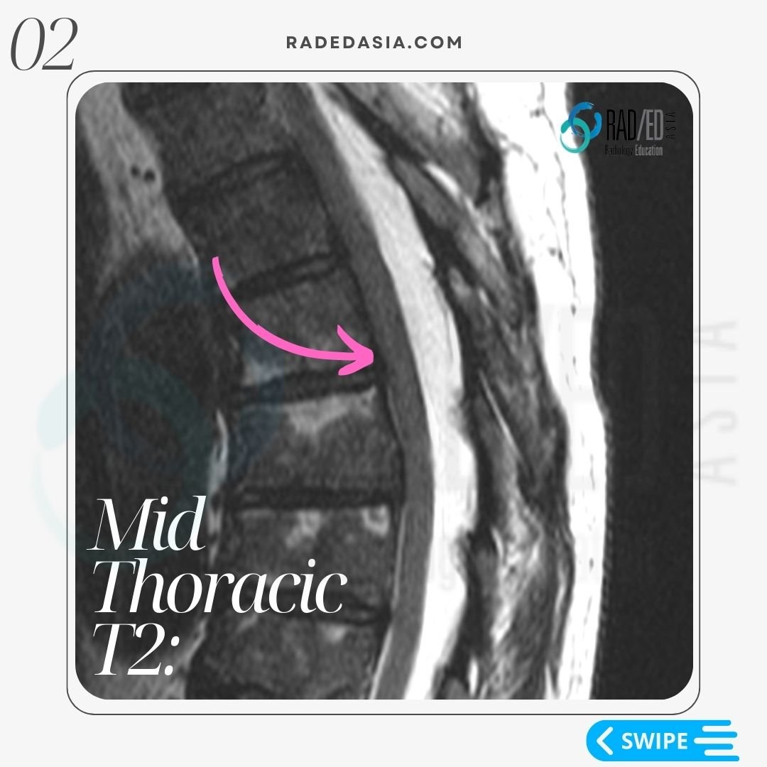
- Thoracic Epidural lipomatosis is not uncommon and has a different appearance on MRI o Lumbar Epidural.
- Unlike the lumbar region, there is no trefoil shape seen.
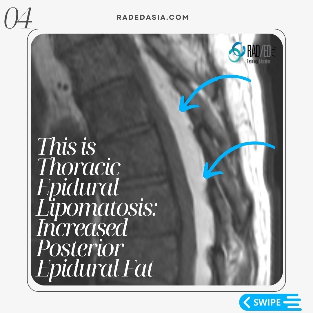
- With thoracic epidural lipomatosis there is excess fat accumulated in the posterior epidural space.
![]()
- Epidural lipomatosis in the thoracic spine has a different appearance to Lumbar Epidural Lipomatosis.
- In the lumbar spine we look for a trefoil appearance of the thecal sac.
- However in the thoracic spine we look for Increased epidural fat.
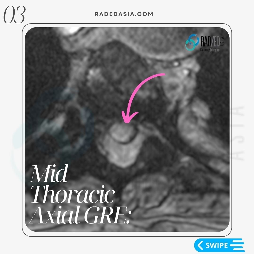
- The fat accumulation is posterior and lateral.
- This results in the Cord being displaced anteriorly and thecal sac effaced.
- There is no trefoil appearance.
![]()
The diagnosis of thoracic epidural lipomatosis on MRI is made by looking for:
- Posterior epidural fat measuring >6mm in AP diameter.
- Effacement of the thecal sac.
- Anterior displacement of the cord.
![]()


Spinal epidural lipomatosis is an excessive accumulation of non-encapsulated fat in the epidural space of the spine.
![]()
Some risk factors for developing spinal epidural lipomatosis include excessive exogenous or endogenous steroids, spinal surgery, and obesity.
The characteristic MRI finding in lumbar spinal epidural lipomatosis is a marked increase in fat in the epidural space, resulting in the characteristic trefoil shape of the compressed thecal sac.
![]()
In the thoracic spine, there is no trefoil shape seen. Instead, there is increased epidural fat accumulation posterior and lateral to the cord, resulting in displacement of the cord anteriorly and effacement of the thecal sac.
![]()
The trefoil shape in lumbar epidural lipomatosis refers to a three-lobed structure arising from a common center, caused by the compressed thecal sac, attached to the dentate ligaments.
![]()
Yes, posterior epidural fat measuring >6mm in AP diameter is a specific measurement used to diagnose thoracic epidural lipomatosis, in addition to the effacement of the thecal sac and anterior displacement of the cord.