
SYNOVITIS MRI FINDINGS: WHAT DOES IT LOOK LIKE
Synovitis on MRI, like the many-faced God in Game of Thrones, can have many appearances. However, like we saw in the article Tendinosis: Same Same but Different once you recognize the different appearances, it’s the same pattern for each joint.
Synovitis is usually generalized and associated with an effusion but can also be localized to a portion of the joint and without an effusion. It’s not possible to differentiate inflammatory from infective synovitis and contrast won’t help as all forms of synovitis will enhance.
We are going to look at the common appearances in this section.![]()
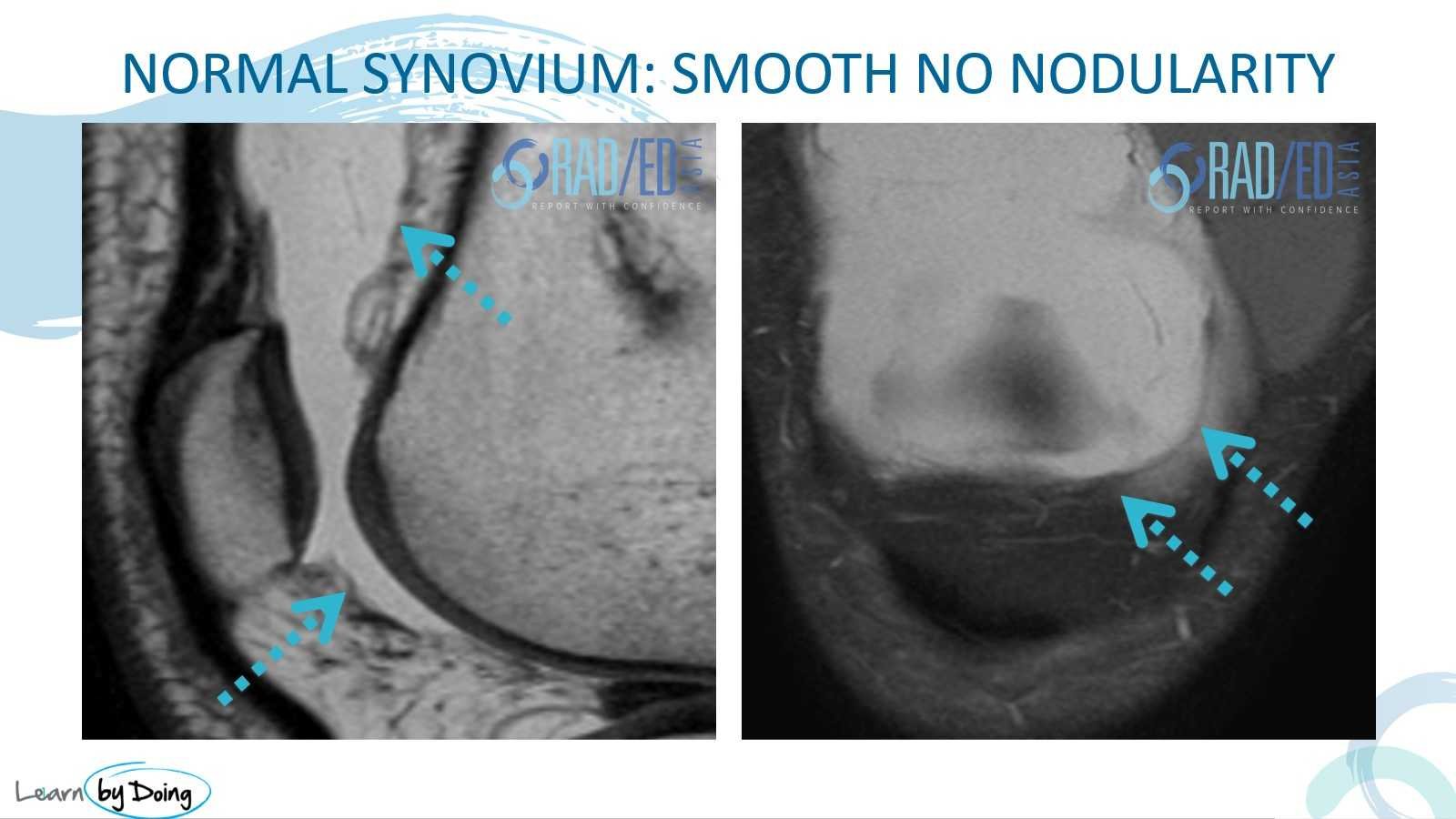
- Normally you cant differentiate synovium from capsule and what we see is a thin low signal line (Blue arrow).
- This is best seen on T2/PD scans.
![]()
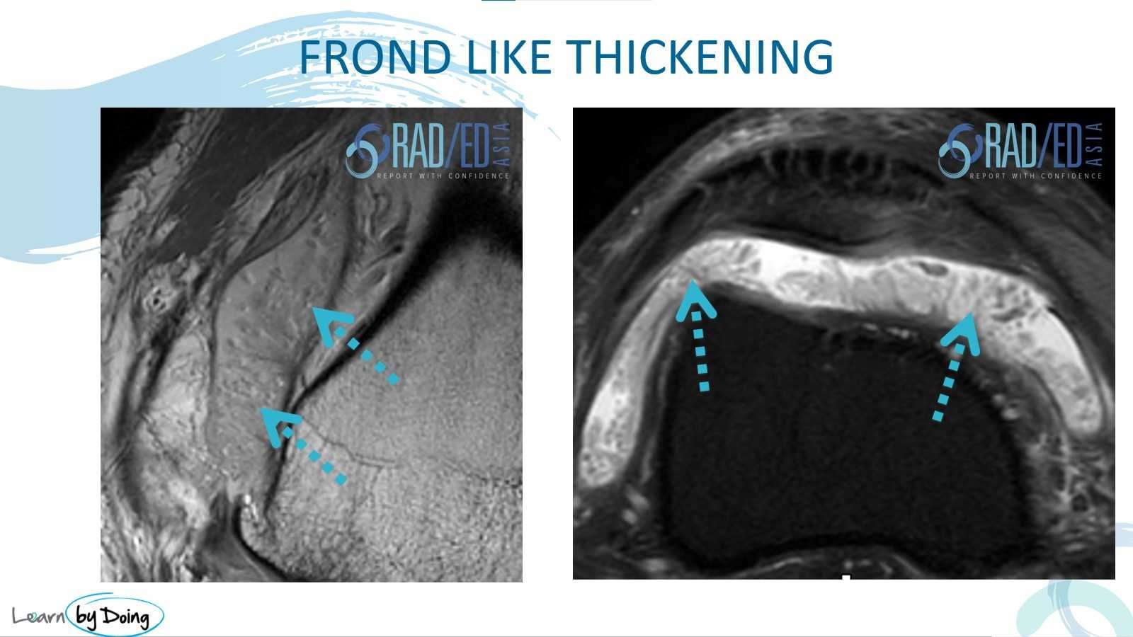
- This is the most common appearance with a thin, fibrillary type appearance that is intermediate in signal on T2 and PD.
- Much easier to see on T2/ PD than on T1.
![]()
- Nodular synovitis has a much thicker appearance than the frond like synovitis.
- It can either be elongated or rounded and nodular.
- The presence of nodular synovitis should raise the possibility of PVNS.

![]()
This is more extensive synovitis that has a more solid appearance.

![]()
- This is similar to the sheet like appearance and lines the capsule of the joint.
- This is type is more commonly seen post ACL repair. and is a type of arthrofibrosis.
- Usually low signal on PD/T2.

![]()

Image above: The 2 images demonstrate intermediate signal from synovitis lying posterior to the meniscus. Often there is an associated meniscal tear (2nd image) which causes the synovitis. When related to a tear, it can be localised just to that area.![]()