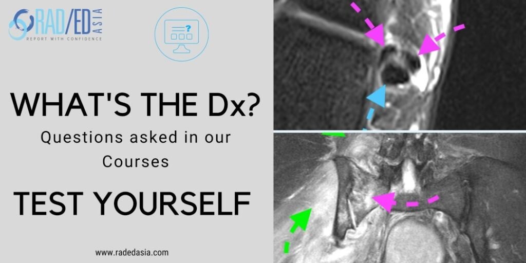
tibialis posterior tendinitis tendinopathy tear mri radiology

The tibialis posterior tendon is enlarged and increased in signal.

TIBIALIS POSTERIOR TENDINOPATHY AND PARTIAL TEAR.

  • Tibialis posterior tendinopathy and delamination / partial tears.


  • The normal tibialis posterior tendon should be uniformly low signal.


  • Enlargement and or increased intermediate signal indicates tibialis posterior tendinopathy / tendinosis.


  • Tibialis posterior tendon tears are indicated by the presence of high signal which is more fluid in appearance.

If your Browser is blocking the video, Please view it on our YouTube Channel HERE.

Learn more about this condition and how to best report it in more detail in our
Guided ANKLE Mini Fellowship.


More by clicking on the images below.

mri periosteum callus periosteal new bone radedasia radiology education
ANKLE
mri achilles tendon abnormalities ankle radiology
ANKLE

#tibialisposterior #tibialisposteriortear #radiology #radedasia #mri #anklemri #msk #mriankle #mskmri #radiologyeducation #radiologycases #radiologist #radiologystudent #radiologycme #radiologycpd #medicalimaging #imaging #radcme #rheumatology #arthritis #rheumatologist #sportsmed #orthopaedic #physio #physiotherapist

#radedasia #mri #mskmri #radiología

Este sitio está destinado únicamente a profesionales médicos. El uso de este sitio se rige por nuestra Términos de servicio y Declaracion de privacidad que se puede encontrar haciendo clic en los enlaces. Acepte antes de continuar con el sitio web.
error: Clic derecho no disponible.