Video de Youtube

subacromial subdeltoid bursa bursitis msk mri radiology radedasia

SUBACROMIAL SUBDELTOID BURSITIS MRI RADIOLOGY (VIDEO)

SUBACROMIAL SUBDELTOID BURSITIS MRI RADIOLOGY DISCUSSION Subacromial subdeltoid (SASD) bursitis on MRI can have a number of appearances. In this case we have a nodular type of synovitis which should raise the possibility of an inflammatory arthritis as a cause (Rheumatoid Arthritis in this case). WHAT DO WE SEE ON THE IMAGE? Fluid in the …

SUBACROMIAL SUBDELTOID BURSITIS MRI RADIOLOGY (VIDEO) Leer más »

ligaments attached to the coracoid process mri shoulder

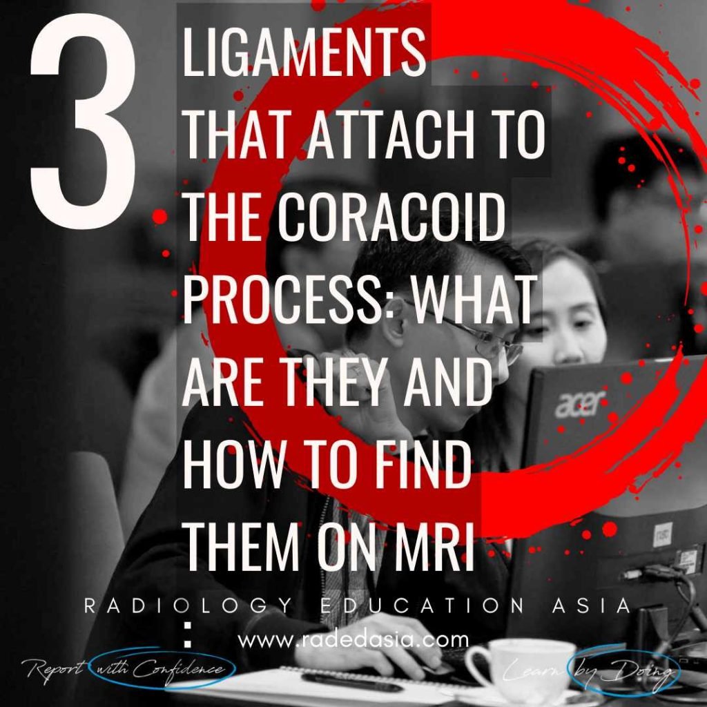
3 LIGAMENTS ATTACHED TO THE CORACOID PROCESS: WHAT ARE THEY AND HOW TO FIND THEM ON MRI (VIDEO)

3 LIGAMENTS ATTACHED TO THE CORACOID PROCESS 3 LIGAMENTS ATTACHED TO THE CORACOID PROCESS: There are three ligaments that attach to the coracoid process of the shoulder. When reporting a shoulder MRI they should be part of the overall assessment. So what are they and how do you find them? See the brief video below. …

3 LIGAMENTS ATTACHED TO THE CORACOID PROCESS: WHAT ARE THEY AND HOW TO FIND THEM ON MRI (VIDEO) Leer más »

ligament retinacula scar mri radiology education asia radedasia

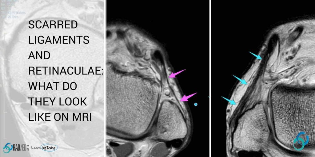
MRI RETINACULA SCAR: SCARRED LIGAMENTS AND RETINACULA: WHAT DOES MRI SCAR TISSUE LOOK LIKE? (VIDEO)

LIGAMENT AND RETINACULA HEALING: WHAT DOES SCAR TISSUE LOOK LIKE ON MRI NEED TO KNOW PATHOLOGY What happens to a ligament when it is torn and injured? This is a brief edited summary from an article on the pathology of scar formation ( “Scar formation and ligament healing” link below).   “After an injury… hemostasis …

MRI RETINACULA SCAR: SCARRED LIGAMENTS AND RETINACULA: WHAT DOES MRI SCAR TISSUE LOOK LIKE? (VIDEO) Leer más »

mri ankle foot ligament short plantar

MRI PLANTAR LIGAMENT SHORT PLANTAR LIGAMENT ANKLE FOOT AND TOE (VIDEO)

MRI PLANTAR LIGAMENT SHORT PLANTAR LIGAMENT WHAT'S THE STRUCTURE The Short Plantar Ligament. WHAT ARE THE FINDINGS ANATOMY OF THE SHORT PLANTAR LIGAMENT. 1. Extends from the calcaneus (Green star) to the cuboid (Yellow star). 2. It is distal and separate to long plantar ligament. 3. Has a fan shaped appearance. MRI PLANTAR LIGAMENT SHORT …

MRI PLANTAR LIGAMENT SHORT PLANTAR LIGAMENT ANKLE FOOT AND TOE (VIDEO) Leer más »

subcortical subchondral fracture knee tibial plateau mri insufficiency

SUBCORTICAL SUBCHONDRAL FRACTURE KNEE MRI TIBIAL PLATEAU (VIDEO)

SUBCORTICAL SUBCHONDRAL FRACTURE KNEE MRI TIBIAL PLATEAU DISCUSSION Subchondral or subcortical fractures of the knee can occur in the femoral condyles or tibial plateau.   The key to the diagnosis is seeing a linear low signal line adjacent to and paralleling the cortex without any cortical defect or break in the acute phase.     There …

SUBCORTICAL SUBCHONDRAL FRACTURE KNEE MRI TIBIAL PLATEAU (VIDEO) Leer más »

spine mri msk online radiology courses radedasia

LUMBAR SPINE ENHANCEMENT MRI ONLINE RADIOLOGY COURSES: KEY POINTS FROM OUR SPINE MRI COURSES

KEY POINTS FROM OUR SPINE MRI COURSES Spine MRI Online Radiology Course: Normal Spinal Enhancement Another three quick images with some basic key points from our online spine MRI courses. Here we look at areas of normal enhancement that can be mistaken for pathology: Normal enhancement of the anterior epidural plexus which can be mistaken …

LUMBAR SPINE ENHANCEMENT MRI ONLINE RADIOLOGY COURSES: KEY POINTS FROM OUR SPINE MRI COURSES Leer más »

tibialis posterior tendinitis tendinopathy tear mri radiology

TIBIALIS POSTERIOR TENDINITIS TENDINOPATHY TEARS RADIOLOGY MRI (VIDEO)

TIBIALIS POSTERIOR TENDINITIS TENDINOPATHY TEARS RADIOLOGY MRI WHAT ARE THE FINDINGS The tibialis posterior tendon is enlarged and increased in signal. DIAGNOSIS & DISCUSSION TIBIALIS POSTERIOR TENDINOPATHY AND PARTIAL TEAR. Tibialis posterior tendinopathy and delamination / partial tears. The normal tibialis posterior tendon should be uniformly low signal. Enlargement and or increased intermediate signal indicates …

TIBIALIS POSTERIOR TENDINITIS TENDINOPATHY TEARS RADIOLOGY MRI (VIDEO) Leer más »

opll spine ossification posterior longitudinal ligament ct mri xray radiology (1)

OPLL SPINE OSSIFICATION POSTERIOR LONGITUDINAL LIGAMENT CT (VIDEO)

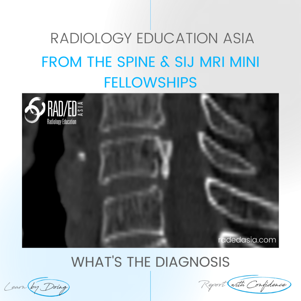
OPLL SPINE OSSIFICATION POSTERIOR LONGITUDINAL LIGAMENT CT DISCUSSION ON OPLL SPINE CT There is localised, linear ossification present at the posterior margin of the C6 vertebra.   This has a typical appearance of OPLL (Ossification of the Posterior Longitudinal Ligament).   The cervical spine is the most common location.   It is important to follow …

OPLL SPINE OSSIFICATION POSTERIOR LONGITUDINAL LIGAMENT CT (VIDEO) Leer más »

ischiofemoral impingement radiology mri hip

ISCHIOFEMORAL IMPINGEMENT RADIOLOGY HIP MRI (VIDEO)

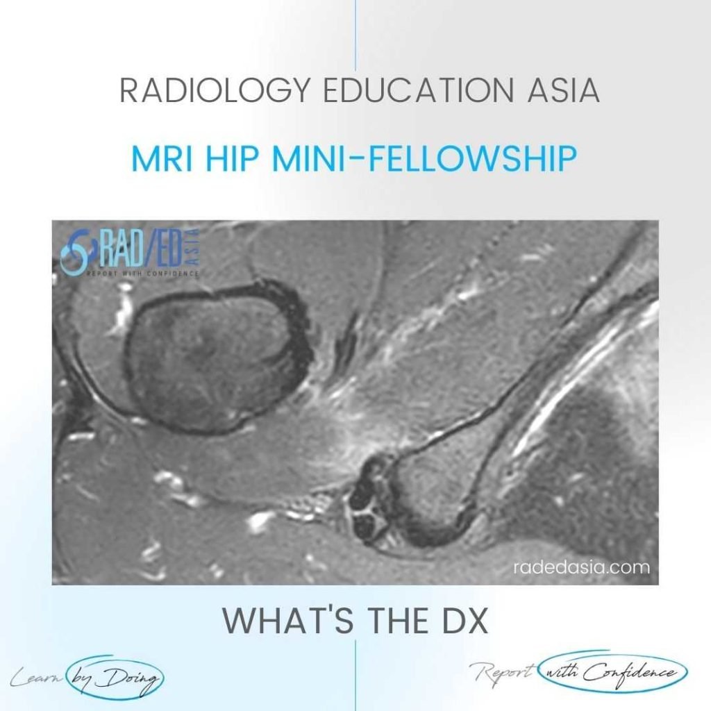
ISCHIOFEMORAL IMPINGEMENT HIP MRI RADIOLOGY ISCHIOFEMORAL IMPINGEMENT: WHY DOES IT OCCUR Ischiofemoral impingement is a result of the quadratus femoris muscle being compressed between the ischial tuberosity and the posterior femur resulting initially in quadratus femoris oedema but can progress to tears and severe atrophy. ISCHIOFEMORAL IMPINGEMENT: WHAT ARE THE FINDINGS Initially there is oedema …

ISCHIOFEMORAL IMPINGEMENT RADIOLOGY HIP MRI (VIDEO) Leer más »

Este sitio está destinado únicamente a profesionales médicos. El uso de este sitio se rige por nuestra Términos de servicio y Declaracion de privacidad que se puede encontrar haciendo clic en los enlaces. Acepte antes de continuar con el sitio web.
error: Clic derecho no disponible.