This site is intended for Medical Professions only. Use of this site is governed by our Terms of Service and Privacy Statement which can be found by clicking on the links. Please accept before proceeding to the website.
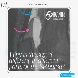
Specific assessment on MRI of knee fat pads for impingement is important. The three fat pads are the suprapatellar or quadriceps fat pad, the supratrochlear fat pad, and the infrapatellar fat pad. Today’s focus is on understanding the suprapatellar fat pad, also known as the quadriceps fat pad, and knowing its location, normal appearance and imaging findings of impingement and how to report Suprapatellar Fat Pad impingement on MRI. The findings associated with suprapatellar fat pad impingement are consistent across the different fat pads, so you can apply these findings to other fat pads as well.
![]()
Read this in conjunction with viewing the video.
Normal Quadriceps or Suprapatellar Fat Pad MRI Anatomy (Video Timestamp 00:33):
Let’s begin with how the normal suprapatellar fat pad appears. It’s a triangular structure, identified behind the quadriceps tendon. On MRI, particularly on the proton density (PD) images, this area is filled with fat, showing high signal intensity. The posterior margin of this fat pad typically aligns with the suprapatellar bursa and should present as either flat or concave. Knowing these normal features is critical to identifying abnormalities.
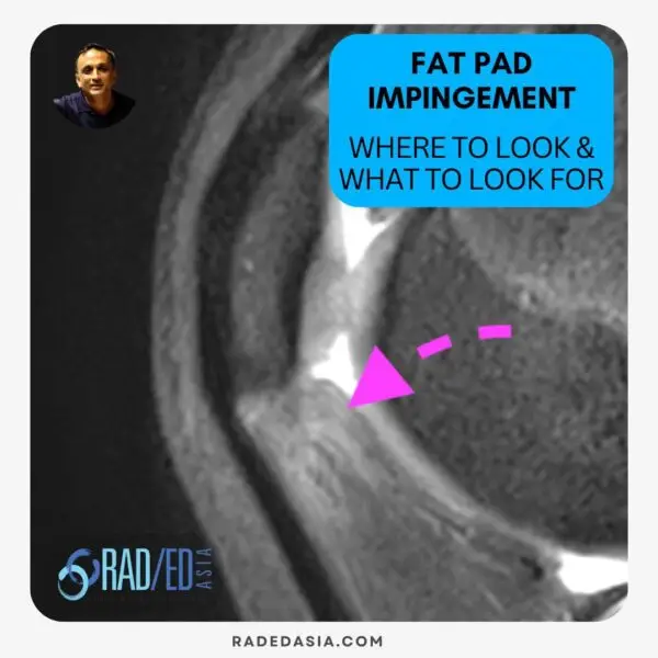
What to Look for: Identifying Abnormal Findings (Video Timestamp 01:29):
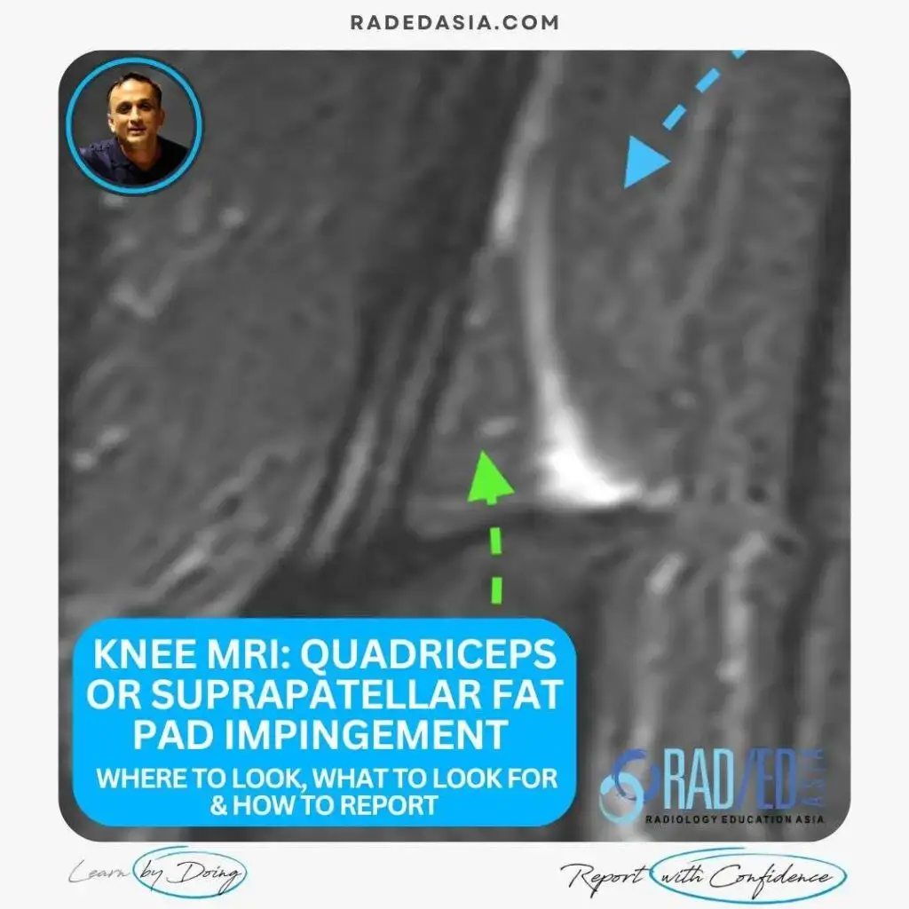
What happens when there are abnormal changes? Impingement may cause the suprapatellar fat pad to show increased signal intensity on MRI. This contrasts with the normal appearance where the fat pad is saturated and darker compared to the bright signal of impingement. Another key indicator is the changing shape of the posterior margin, often transitioning to a convex shape due to oedema pushing it outwards. Similar findings can be observed in other fat pads, including the supratrochlear and infrapatellar fat pads, when increased signal is present.
What to Report (Video Timestamp 02:21):
Reporting these findings of impingement is straightforward. Highlight any increased signal in the suprapatellar fat pad, mentioning any expansion or convexity at the posterior margin, and note if there’s any indentation on the supratrochlear fat pad.
![]()
Article: “AJR Quadriceps Fat Pad Signal Intensity and Enlargement on MRI”, Read HERE
![]()
For those interested in becoming much more confident in assessing and reporting Knee MRI, consider joining your colleagues from around the world in our guided Online Knee MRI Mini Fellowship to recognize normal structures, pinpoint abnormalities, and accurately report findings, boosting your diagnostic confidence.
More by clicking on the images below.
Additionally, you can join our WhatsApp community group at this link https://bit.ly/radedasiacommunity where we regularly post educational material that’s quick and easy to learn.
![]()