Sagittal PD MRI of the knee showing toothbrush-like irregularity of the cartilage surface consistent with chondral fibrillation

Stay tuned on new Fellowships and learnings

KNEE MRI: CHONDRAL FIBRILLATION – WHAT TO LOOK FOR (VIDEO)

KNEE MRI: CHONDRAL FIBRILLATION – WHAT TO LOOK FOR

What is chondral fibrillation?

Chondral fibrillation is the earliest visible change in damaged articular cartilage, appearing after an initial phase of cartilage softening. On MRI it shows up as an irregular, roughened cartilage surface in place of the normal smooth, sharp margin — and the knee, where cartilage is thick, is the easiest joint in which to see it.

If you find the video helpful, please subscribe to the channel.

THE SPECTRUM OF CARTILAGE CHANGE

Knee cartilage can show a number of different types of change, progressing along a spectrum:

  • Softening — the earliest change of all, preceding any visible surface irregularity.
  • Fibrillation — the earliest chondral change actually seen on imaging, an irregularity of the cartilage surface/margin.
  • Fissuring — fibrillation progresses to defined clefts within the cartilage.
  • Full-thickness chondral loss — the end stage of this progression.
HOW TO RECOGNISE FIBRILLATION ON MRI

On the sagittal PD sequence, normal cartilage shows intermediate signal with a nice sharp, crisp margin. Where fibrillation is present, that margin is lost:

  • The cartilage surface takes on a characteristic “toothbrush” appearance — multiple small, bristle-like irregularities along the surface, rather than a single smooth line.
  • This is a surface-level change: the fibrillation itself has not yet progressed to a discrete fissure or to full-thickness loss.
WHERE FIBRILLATION IS SEEN
  • Chondral fibrillation is not uncommon in the knee, and the knee is actually the easiest joint in which to see it.
  • This is because knee cartilage is comparatively thick, making surface irregularity easier to resolve on MRI.
  • It can occur at the patellofemoral joint, or at the medial or lateral compartment.

Fibrillation can also occasionally be seen in other joints, but it is harder to identify because cartilage thickness elsewhere is much less than in the knee.

KEY POINT
  • Chondral fibrillation is the earliest chondral change seen on imaging, occurring after an initial phase of cartilage softening.
  • It appears as irregularity of the cartilage surface/margin — on sagittal PD imaging this produces a “toothbrush” appearance in place of the normal sharp, crisp margin.
  • Untreated, fibrillation can progress to fissuring and eventually full-thickness chondral loss.
  • It is easiest to identify in the knee, where cartilage is thick, occurring at the patellofemoral joint or the medial/lateral compartment; it is harder to see in other, thinner-cartilage joints.
TEST YOURSELF ON SOME COMMON & FREQUENTLY ASKED QUESTIONS

Fibrillation, which follows an initial phase of cartilage softening. It is the earliest change that is actually visible as surface irregularity of the cartilage.

Normal cartilage has intermediate signal and a sharp, crisp margin. With fibrillation, that margin becomes irregular, producing a characteristic “toothbrush” appearance on the surface.

Knee cartilage is comparatively thick, which makes surface irregularity easier to resolve on MRI than in joints with thinner cartilage.

It progresses to fissuring, and eventually to full-thickness chondral loss.

KNEE MRI CHONDRAL FIBRILLATION – VIEW VIDEO

If you wish to watch the video in language other than English, please view it on our YouTube Channel HERE

Our CPD & Learning Partners

OUR POPULAR POSTS ON KNEE FRACTURES: CLICK ON THE IMAGES BELOW

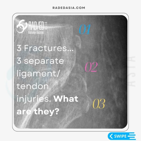
MRI evaluation of ACL-associated second fractures, highlighting key secondary osseous injuries associated with anterior cruciate ligament tears.
Anterior Cruciate Ligament
radiology knee segond arcuate lateral fibular fracture xray mri knee radedasia
KNEE

Stay tuned on new Fellowships and learnings

error: Content is protected !!

Join our Newsletter

Stay tuned on new
Mini-Fellowships launches and learnings

Mini-Fellowships

Better Than Conferences

Join our WhatsApp Community

We help radiologists learn
by doing and report with confidence

This site is intended for Medical Professions only. Use of this site is governed by our Terms of Service and Privacy Statement which can be found by clicking on the links. Please accept before proceeding to the website.