MRI arachnoiditis illustrating pathogenesis and normal cauda equina nerve root distribution.

Stay tuned on new Fellowships and learnings

MRI ARACHNOIDITIS: NORMAL NERVE ROOT DISTRIBUTION – WHAT TO LOOK FOR (VIDEO)

NORMAL NERVE ROOT DISTRIBUTION: WHAT TO LOOK FOR

Before you can spot abnormal nerve roots, you need to know what normal looks like

  • This is part 1 of our series on the MRI appearance of arachnoiditis. Arachnoiditis is not rare — it's often thought of only in terms of infection, but it's relatively common in post-operative spines.
  • In this video we cover the pathogenesis of arachnoiditis — why it happens — and the normal distribution of nerve roots in the lumbar spine, since knowing normal is what lets you recognise abnormal.
  • Part 2 of this series covers normal contrast enhancement patterns, which you need to know before interpreting arachnoiditis studies given with contrast.

If you find the video helpful, please subscribe to the channel.

WHAT IS ARACHNOIDITIS AND WHY DOES IT HAPPEN
  • Arachnoiditis is inflammation of the arachnoid membrane, made up of the dura, arachnoid, and pia.
  • These layers are avascular — important to know, since it affects what you'd expect to see with contrast.
  • Acute inflammation, infection, or hemorrhage leads to fibrin and exudate being deposited on the nerve root sheaths within the arachnoid.
  • Over time this produces collagen formation, and the nerve roots start to stick together.
  • Similar collagen change within the subarachnoid space can form septae.
  • Postoperative change is probably the most common cause in many practices; infection is a common cause in other countries.
  • Up to around 20% of post-laminectomy or post-fusion patients develop some form of arachnoiditis.
  • Radiculitis or early septae formation postoperatively is actually quite common, but doesn't always persist — the persistent cases make up that ~20% figure.
  • Surgery doesn't need to go through the dural space to cause it: even extradural surgery can result in arachnoiditis or radiculitis, apparently through changes in vascularity.
  • Other causes include trauma, hemorrhage, and injections into the subarachnoid space (e.g. spinal tap or puncture).
  • This series doesn't cover malignant arachnoiditis specifically, since it isn't a true arachnoiditis.
  • It's tumour cells lining the arachnoid, not an inflammatory or infective process.
NORMAL NERVE ROOT DISTRIBUTION – THE CAUDA EQUINA APPEARANCE

Proximally, at the level of the conus, the nerve roots are centred close together, bunched around it.

The nerve roots start to separate out, becoming progressively further apart from each other as you move distally — this is the cauda equina ("horse's tail") appearance.

The nerve roots are fully separated out, distributed around the margin of the spinal canal. Seeing them bunched together at this level — instead of separated peripherally — is abnormal.

WHAT TO CHECK ON AXIAL IMAGES
  • At the S1 level, the nerve roots lie peripherally, right up against the edge of the thecal sac.
  • There should still be a visible rim of CSF separating the nerve root from the thecal sac margin — not the nerve root stuck directly against the dura.
  • Sagittal images show the nerve roots fanning out — the cauda equina appearance.
  • Axial images are more useful than sagittal for assessing whether a nerve root distribution is normal.
KEY POINT
  • Arachnoiditis is inflammation of the arachnoid membrane (dura, arachnoid, pia) — avascular layers, which matters when later interpreting contrast enhancement.
  • Postoperative change is a common cause; up to around 20% of post-laminectomy or fusion patients develop some form of arachnoiditis, and even extradural surgery can trigger it.
  • Normal nerve roots are bunched centrally near the conus and progressively separate more distally, lying peripherally by S1 — but should still show a rim of CSF separating them from the thecal sac margin.
  • A bunched-up nerve root pattern at S1 is abnormal; axial images are more useful than sagittal for assessing this.
TEST YOURSELF ON SOME COMMON & FREQUENTLY ASKED QUESTIONS
Inflammation of the arachnoid membrane — the dura, arachnoid, and pia — which are avascular layers.

  • Postoperative change is the most common cause in many practices, though infection is more common in some countries.
  • Other causes include trauma, hemorrhage, and injections into the subarachnoid space.

The nerve roots lie peripherally, distributed around the margin of the spinal canal, with a visible rim of CSF separating them from the thecal sac — not bunched together, and not stuck directly against the dura.

If you wish to watch the video in language other than English, please view it on our YouTube Channel HERE

We look at all of these topics in more detail in our Guided SPINE & SIJ Imaging Mini Fellowships.

Click on the image below for more information.

Our CPD & Learning Partners

OTHER POPULAR SPINE REPORTING POSTS: CLICK ON THE IMAGES BELOW

PAIN MANAGEMENT
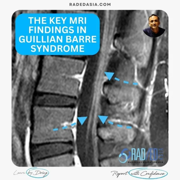
guillain-barre-syndrome-mri-findings-nerve-enhancement-spine-radiology-radedasia
Spanish Blogs

Stay tuned on new Fellowships and learnings

error: Content is protected !!

Join our Newsletter

Stay tuned on new
Mini-Fellowships launches and learnings

Mini-Fellowships

Better Than Conferences

Join our WhatsApp Community

We help radiologists learn
by doing and report with confidence

This site is intended for Medical Professions only. Use of this site is governed by our Terms of Service and Privacy Statement which can be found by clicking on the links. Please accept before proceeding to the website.