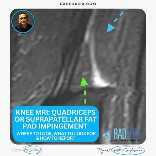
MRI sagittal view of the knee showing the medial gastrocnemius tendon insertion with degenerative findings.

Stay tuned on new Fellowships and learnings

MRI KNEE: MEDIAL GASTROCNEMIUS KNEE TENDON DEGENERATION

FROM TODAY'S REPORTING

MEDIAL GASTROCNEMIUS KNEE TENDON DEGENERATION


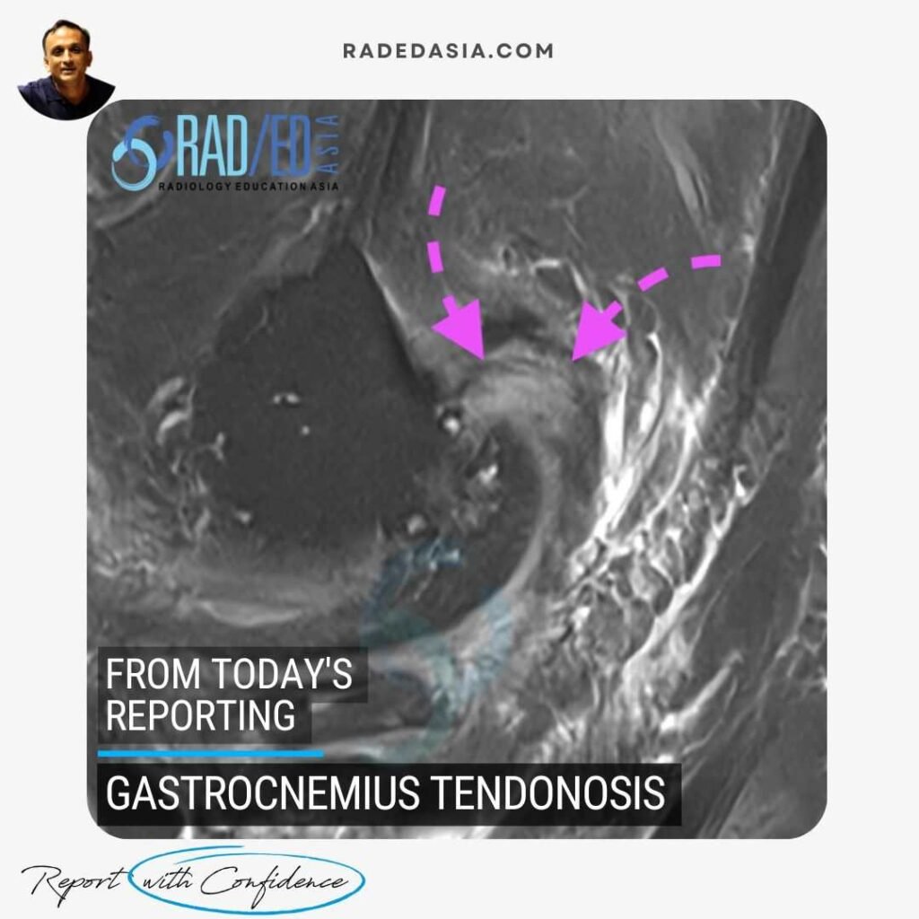
Medial gastrocnemius tendon degeneration and tears can be a source of posterior knee pain. What to look for on MRI? The same things that occur in tendons elsewhere.


MEDIAL GASTROCNEMIUS TENDON DEGENERATION: MRI FINDINGS

  • Tendon thickening or irregularity (Pink arrow) at and adjacent to the femoral condyle insertion.

  • Tendon ill definition.

  • Intermediate increased signal intensity on PDFS or T2FS -weighted images for degeneration (Pink arrow).

  • Higher fluid type  increased signal intensity on PDFS or T2FS -weighted images for tears.

Irregularity of the cortical margin, bone marrow oedema or subcortical cyst formation at the femoral condyle.

TEST YOURSELF ON SOME COMMON & FREQUENTLY ASKED QUESTIONS

Increased signal intensity within the tendon, thickening, potential partial or full-thickness tears, and signs of subcortical cystic change or marrow oedema at the attachment.

Sagittal and axial T2-weighted or proton density fat-saturated sequences.

Our CPD & Learning Partners

OUR POPULAR POSTS ON KNEE MRI: CLICK ON THE IMAGES BELOW

knee mri quadriceps or suprapatellar fat pad impingement radiology
KNEE
acl-pcl-mucoid-degeneration-mri-findings-radedasia
Anterior Cruciate Ligament

Stay tuned on new Fellowships and learnings

error: Content is protected !!

Join our Newsletter

Stay tuned on new
Mini-Fellowships launches and learnings

Mini-Fellowships

Better Than Conferences

Join our WhatsApp Community

We help radiologists learn
by doing and report with confidence

This site is intended for Medical Professions only. Use of this site is governed by our Terms of Service and Privacy Statement which can be found by clicking on the links. Please accept before proceeding to the website.