
A FEW MRI MENISCUS ABNORMALITIES: MENISCUS TEAR, INTRA MENISCAL CYST AND VARIANTS THAT LOOK LIKE TEARS
MRI MENISCUS ABNORMALITIES
A few MRI Meniscus abnormalities such as Meniscus tear, Intra meniscal cyst and variants that look like tears.
WHAT ARE THE FINDINGS?
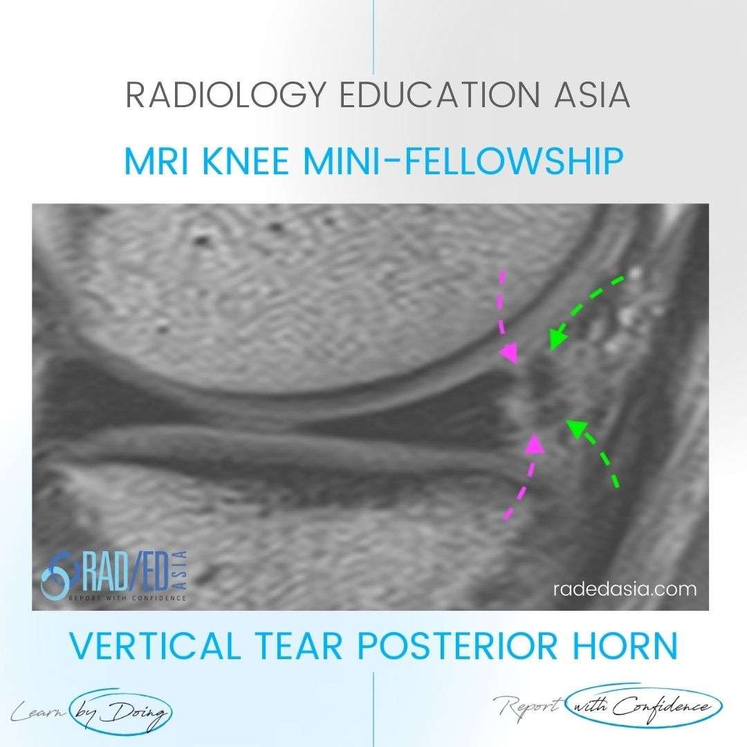
Linear high signal extending from the inferior to superior articular surface (Pink arrows) of the posterior horn medial meniscus.
WHAT'S THE Dx?
VERTICAL (LONGITUDINAL) MENISCUS TEAR POSTERIOR HORN
- The high signal extends to both the superior and inferior articular margins of the meniscus indicating a tear.
- It can be easy to overlook peripheral vertical tears as the torn outer fragment (Green arrows) can be very thin and may not be interpreted correctly as torn meniscus.
- Look for tissue that is the same signal as the parent meniscus and also irregularity of the residual meniscus margin which is torn.


WHAT ARE THE FINDINGS?
Displaced meniscal tissue (Pink arrows) deep to the superficial MCL (Green arrow).
WHAT'S THE Dx?
Flap tear of the medial meniscus body. This is a horizontal tear through the body and the inferior portion has flipped out (Pink arrows) of the joint and lies in the recess between the superficial MCL (Green arrow) and tibia. The residual body (Blue arrow) in the joint is small reflecting loss of meniscus.
Learn more about this condition & How best to report it in more detail in our Online Guided KNEE MRI Mini Fellowship.
More by clicking on the images below.
For all our other current MSK MRI & Spine MRI
Online Guided Mini Fellowships.
Click on the image below for more information.
- Join our WhatsApp RadEdAsia community for regular educational posts at this link: https://bit.ly/radedasiacommunity
- Get our weekly email with all our educational posts: https://bit.ly/whathappendthisweek


