Patella Tendinosis and Tears What to look for on MRI
Tendinosis and Tears of the Patella Tendon on MRI are pretty straightforward and follow the same pattern as tears and tendinosis elsewhere. Here is what to look for on MRI.

1. PATELLA TENDINOSIS
A. Increased PD/PDFS signal but not fluid strength.
B. The tendon may increase in size.
C. Look for spread of inflammation into Hoffa’s Fat Pad or oedema in the inferior pole of the patella at the attachment site.
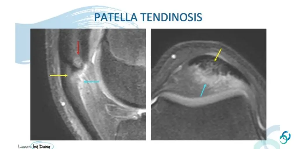
Image Above: Patella tendinosis (Blue arrow). Tendon enlarged and hyperintense (but not fluid type signal).
 Image Above: Patella tendinosis (Yellow arrow). Tendon enlarged and hyperintense (but not fluid type signal). Reactive oedema in patella at attachment (Red arrow) and oedema/inflammatory changes spreading to adjacent Hoffa’s fat (Blue arrow).

2. PARTIAL TEAR
A: Increased PD/PDFS signal in tendon with fluid signal strength.
B: +/- Inflammation in Hoffa’s Fat pad and oedema in patella at insertion site.
 Image Above: Patella tear (Red arrow fluid type signal). Reactive oedema in patella at attachment (Blue arrow) and oedema/ inflammatory changes spreading to adjacent Hoffa’s fat (Yellow arrow).
 Image Above: Patella tear (Blue arrow) demonstrates fluid type T2 signal. Tendon enlarged in keeping with tendinosis.

Our CPD & Learning Partners
- Join our WhatsApp RadEdAsia community for regular educational posts at this link: https://bit.ly/radedasiacommunity
- Get our weekly email with all our educational posts: https://bit.ly/whathappendthisweek


