This site is intended for Medical Professions only. Use of this site is governed by our Terms of Service and Privacy Statement which can be found by clicking on the links. Please accept before proceeding to the website.

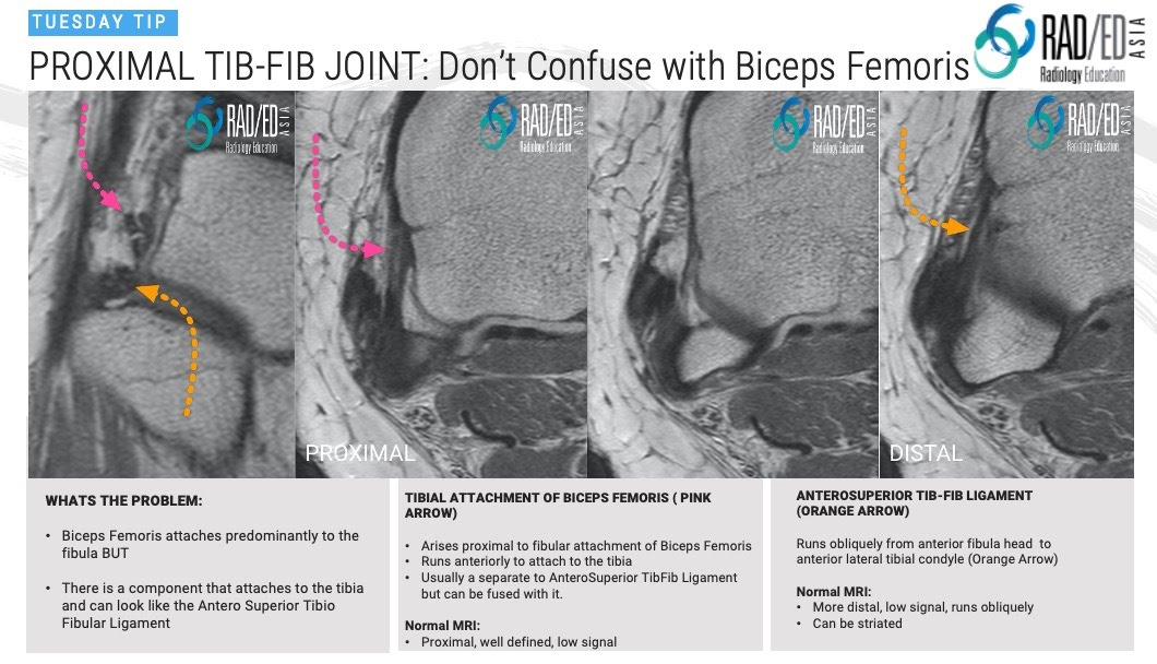
On MRI at the Proximal TibioFibular Joint (PTFJ) one thing that can be confusing is the tibial attachment of the Biceps Femoris Tendon which can be mistaken for the AnteroSuperior TibioFibular Ligament.
![]()
Today’s Tuesday Tip continues with the Proximal TibioFibular Joint. On MRI at the Proximal TibioFibular Joint (PTFJ) one thing that can be confusing is the tibial attachment of the Biceps Femoris Tendon which can be mistaken for the AnteroSuperior TibioFibular Ligament.
The Biceps Femoris tendon attaches predominantly to the fibula head but there is a component that
![]()