mri-subscapularis-tendon-tear-atrophy-fatty-infiltration-biceps-subluxation-dislocation-radedasia

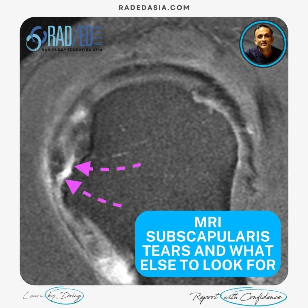
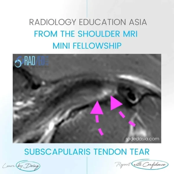
MRI Assessment of the subscapularis tendon is not just about looking for tears. This post looks at the four things that should be assessed. 

  • The MRI appearance of partial and full-thickness tears.
     
  • Then assessing for muscle atrophy and fatty infiltration to determine prognosis from any tendon repair.
     
  • And looking for secondary biceps subluxation or dislocation and understanding the importance of the subscapularis insertion to biceps tendon stability.

mri-subscapularis-tendon-tear-radedasia

  • PDFS axial demonstrates increased fluid type signal at the Subscapularis Tendon insertion in keeping with a deep surface partial tear.


  • Deep surface is where the tears commonly begin.


  • Look for discontinuity of tendon fibres and increased fluid type signal intensity on T2 or PD Fat Sat (T2FS/ PDFS) images.

mri-subscapularis-tear-shoulder-radiology-radedasia


  • T2FS sagittal demonstrates increased fluid type signal at the Subscapularis Tendon insertion in keeping with a deep surface tear.


  • I find the sagittal scans much better to spot tears before I look at the axial scans.


  • Look for discontinuity of tendon fibres and increased fluid type signal intensity on T2 or PD Fat Sat (T2FS/ PDFS) images.

mri-subscapularis-tendon-tear-atrophy-fatty-infiltration-radedasia


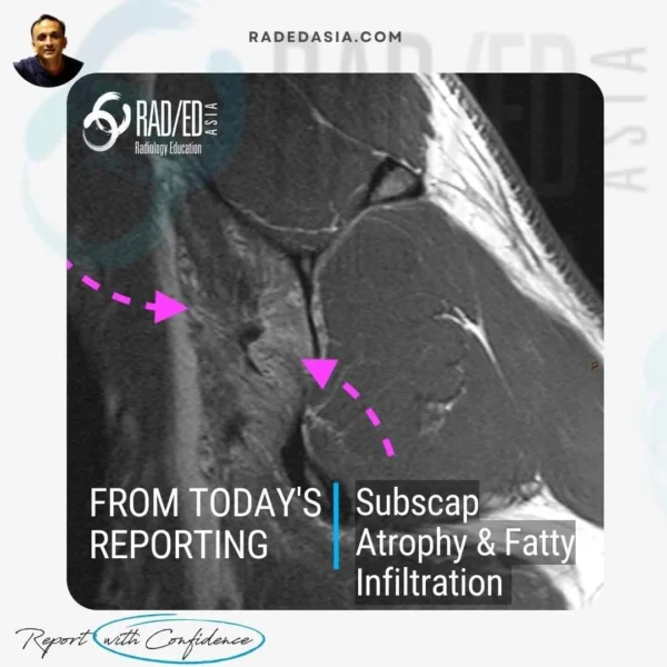
Muscle atrophy and fatty infiltration are two important findings to look for in any rotator cuff tendon tear.

  • MUSCLE ATROPHY,
    1. Look for reduction in size of the muscle and reduction in muscle fibres.
    2. For subscapularis this is best assessed on Sagittal T1 pr PD scans.
    3. Do not assess on axial or Fat saturated images as its harder.
    4. Extent of muscle atrophy important to report as it affects prognosis of tendon repairs.


  • FATTY INFILTRATION,
    1. Occurs after atrophy commences
    2. Fat cells infiltrate the muscle belly.
    3. Look for increased T1 or PD signal in the muscle.
    4. For subscapularis this is best assessed on Sagittal scans.
    5. Do not assess on axial or Fat saturated images. Fat saturation will remove the fat signal and you cant assess the amount of fatty infiltration.
    6. Extent of fatty infiltration important to report as it affects prognosis of tendon repairs.

mri-subscapularis-tendon-tear-biceps-subluxation-dislocation-radedasia

  • Axial scan. Biceps tendon (Both pink arrows) medially subluxed and perched on top of the medial margin of the bicipital groove (Green arrow).

  • This was a result of a subscapularis tear (not shown). See the next tab that explains how a subscapularis tear and biceps subluxation/dislocation are connected.

  • Increased intermediate signal in the biceps tendon from tendonosis.

mri-subscapularis-tendon-tear-anatomy-radedasia

  • Wonderful histological image of the relationship of subscapularis attachment to the biceps tendon in the bicipital groove where subscapularis forms a medial wall and prevents medial movement of biceps.

  • This explains why a tear of subscap can result in medial subluxation/dislocation of the biceps tendon.


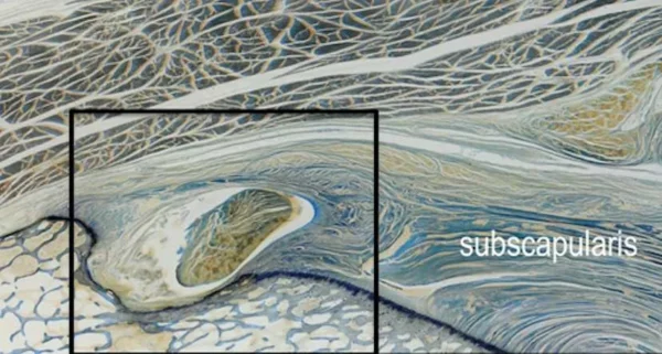
Image from: Tamborrini G et al. The Rotator Interval Ultrasound Int Open 2017; 3: E107–E116.

Our CPD & Learning Partners

Learn more about this condition & How best to report it in more detail in our Online Guided SHOULDER MRI Mini Fellowship.

More by clicking on the images below.

For all our other current MSK MRI & Spine MRI
Online Guided Mini Fellowships.

Click on the image below for more information.

#radedasia #mri #mskmri #radiology

This site is intended for Medical Professionals only. Use of this site is governed by our Terms of Service and Privacy Statement which can be found by clicking on the links. Please accept before proceeding to the website.
error: Right Click not available.