This site is intended for Medical Professions only. Use of this site is governed by our Terms of Service and Privacy Statement which can be found by clicking on the links. Please accept before proceeding to the website.

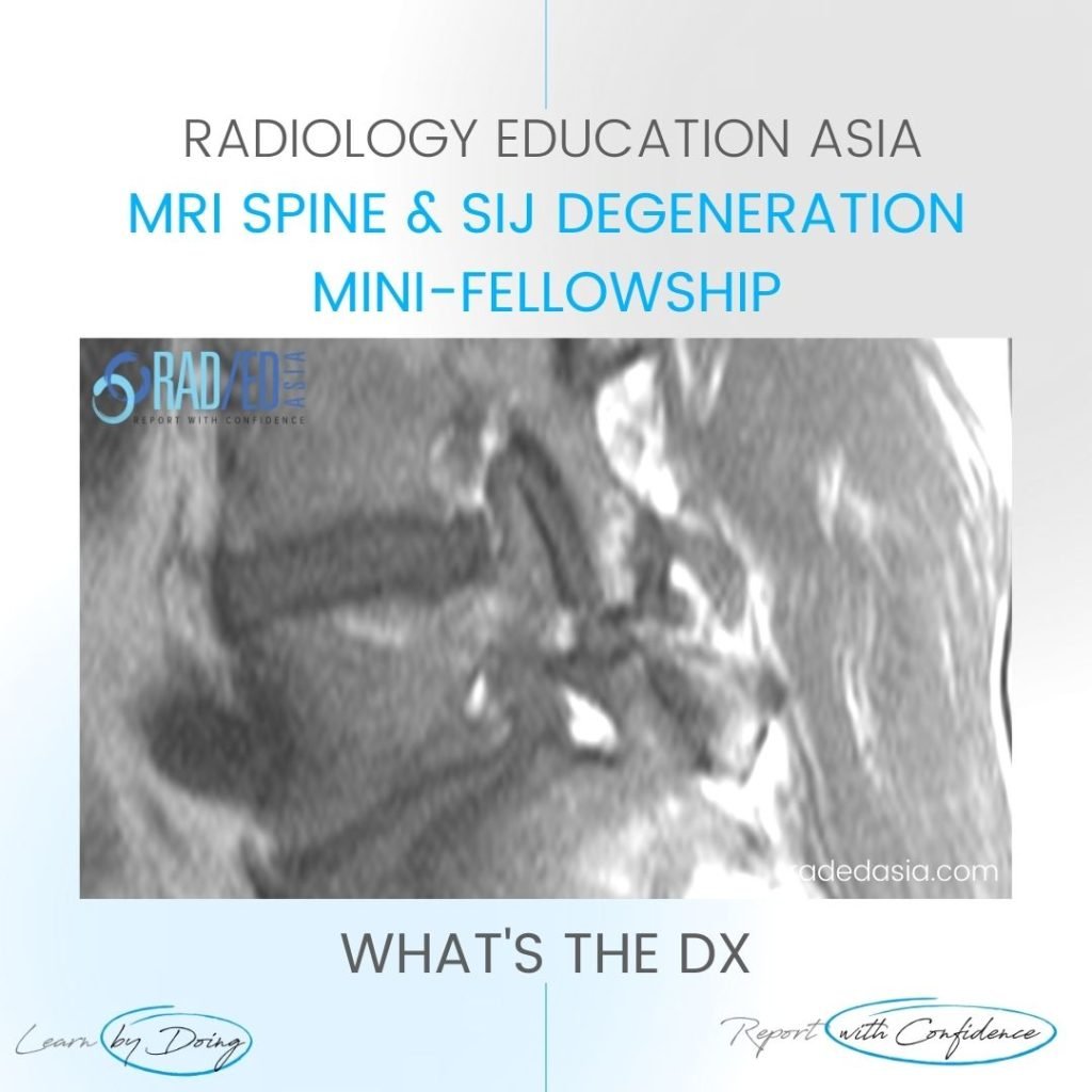
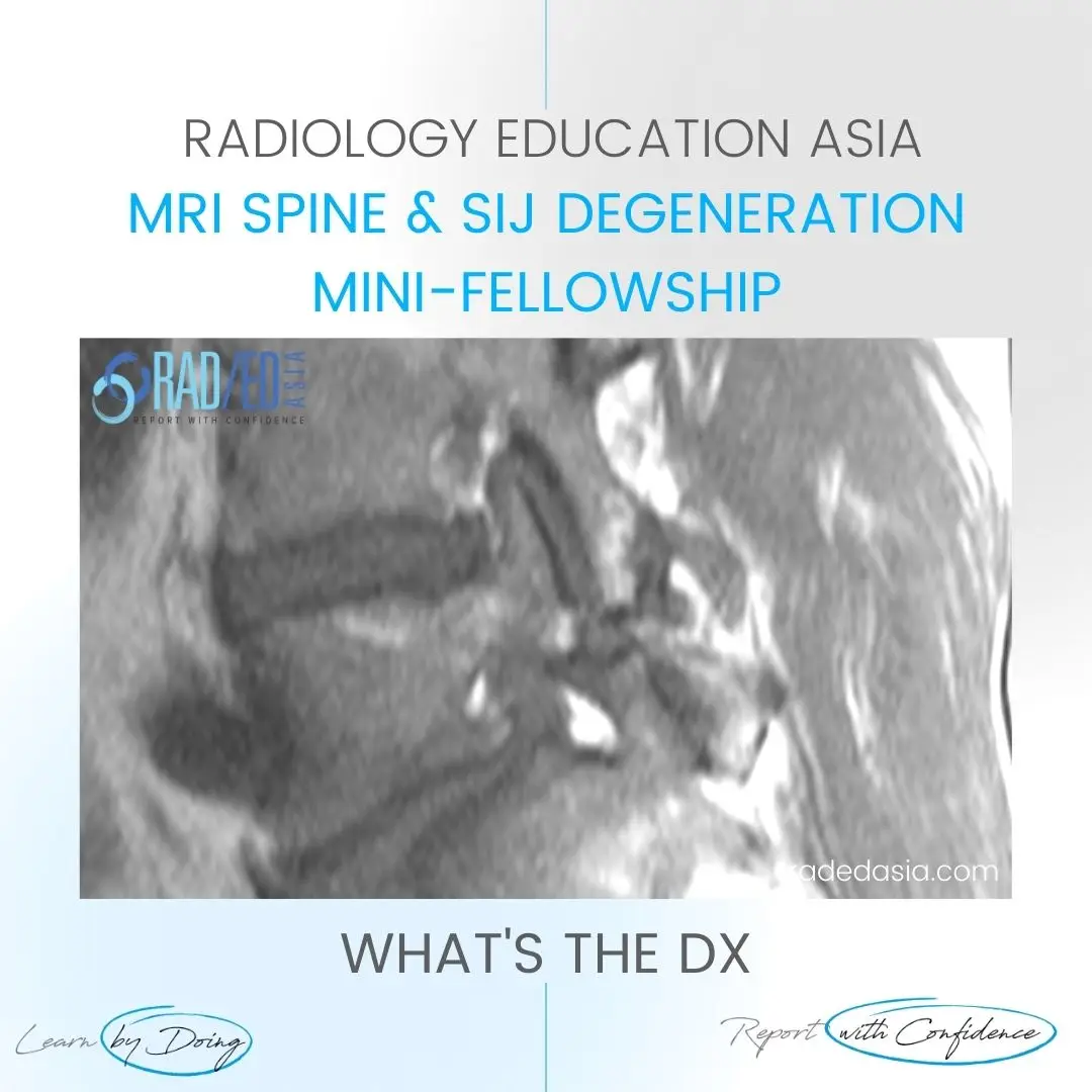
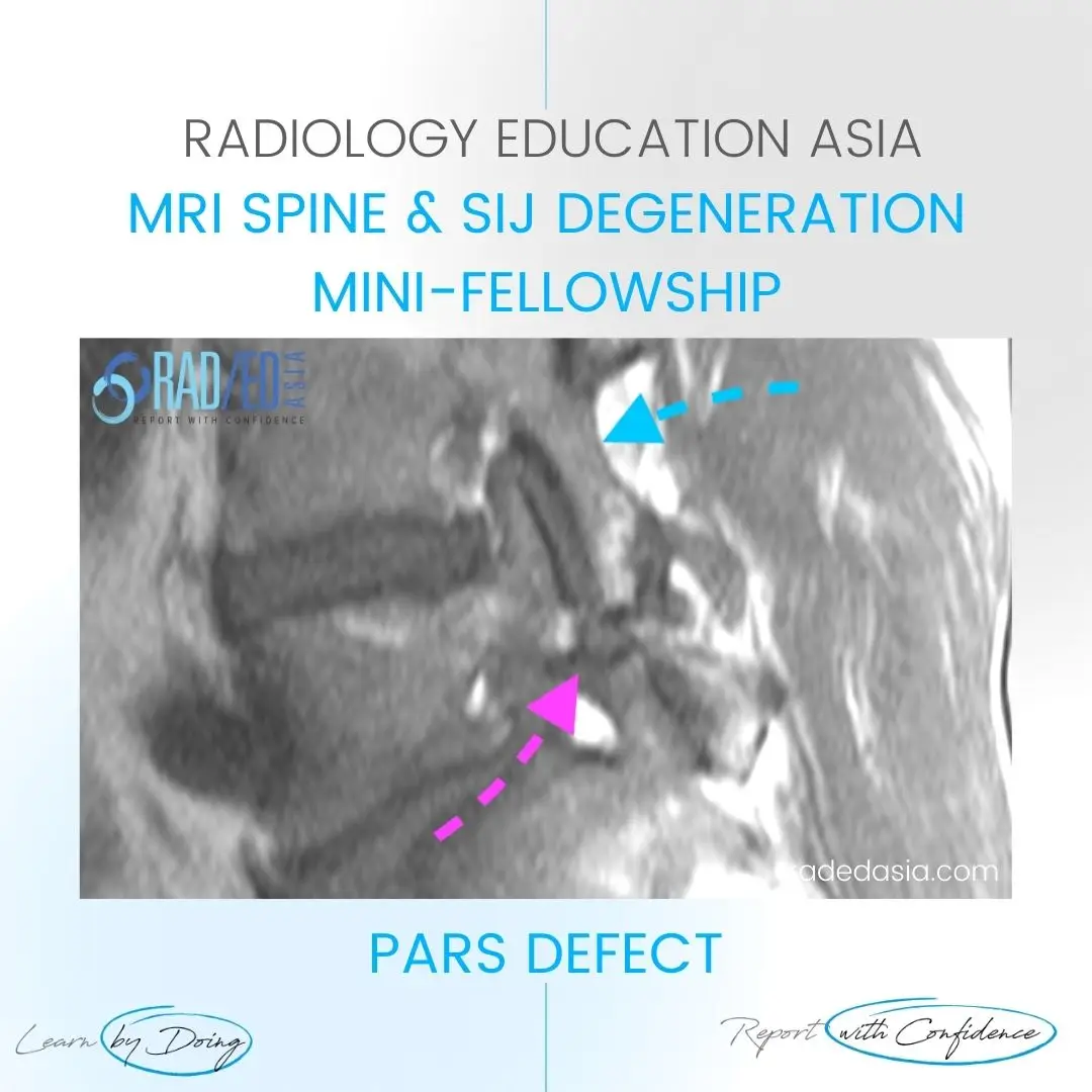
PARS DEFECT


#parsdefect #radiology #radedasia #mri #spinemri #mskmri #mrispine #radiologyeducation #radiologycases #radiologist #radiologycme #radiologycpd #medicalimaging #imaging #spinedegeneration #rheumatology #arthritis #rheumatologist #degenerativedisease #orthopaedic #painphysician #chiropractic #chiropracter #physiotherapy #sportsmed #orthopaedic #mskmri #parsdefect #physicaltherapy #physicaltherapist

Stay tuned on new
Mini-Fellowships launches and learnings
This site is intended for Medical Professions only. Use of this site is governed by our Terms of Service and Privacy Statement which can be found by clicking on the links. Please accept before proceeding to the website.