epidural lipomatosis radiology mri

Epidural lipomatosis is not uncommon to see and is an important diagnosis to make as it can be symptomatic.

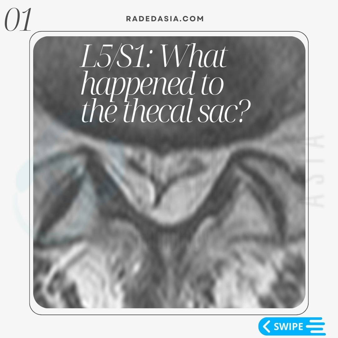
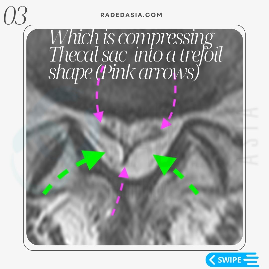
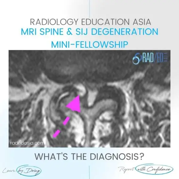
Marked increase in fat in the epidural space (Green arrows) with the thecal sac compressed into a characteristic trefoil shape (Pink arrow).

The trefoil appearance of the thecal is a very characteristic appearance in the lumbar spine with epidural lipomatosis.

However this appearance is not seen in the thoracic spine where there will just be excess epidural fat.

In the cervical spine epidural lipomatosis is not seen.

  • The trefoil appearance refers to a three lobed structure arising from a common centre.

  • The sac compresses into that shape because Dentate ligaments attach to the 3 margins preventing the sites of attachment from being compressed.

For the characteristic MRI appearance of Lumbar Epidural lipomatosis, see the images and explanation below.

We look at all of these topics in more detail in our SPINE MRI Mini Fellowships.

Click on the image below for more information.

Our upcoming onsite Guided MSK MRI Mini Fellowships.

Click on the image below for more information.

spine mri incidental findings parotid mass
SPINE
facet synovial cyst radiology lumbar spine mri
SPINE

#epidurallipomatosis #radiology #radedasia #mri #spinemri #radiologyeducation #radiologycases #radiologist #radiologycme #radiologycpd #medicalimaging #imaging #radcme #rheumatology #arthritis #rheumatologist #orthopaedic #painphysician #chiropractic #chiropracter #physiotherapy #sportsmed #orthopaedic #spinedegeneration #epidurallipomatosis

#radedasia #mri #mskmri #radiology

This site is intended for Medical Professionals only. Use of this site is governed by our Terms of Service and Privacy Statement which can be found by clicking on the links. Please accept before proceeding to the website.
error: Right Click not available.