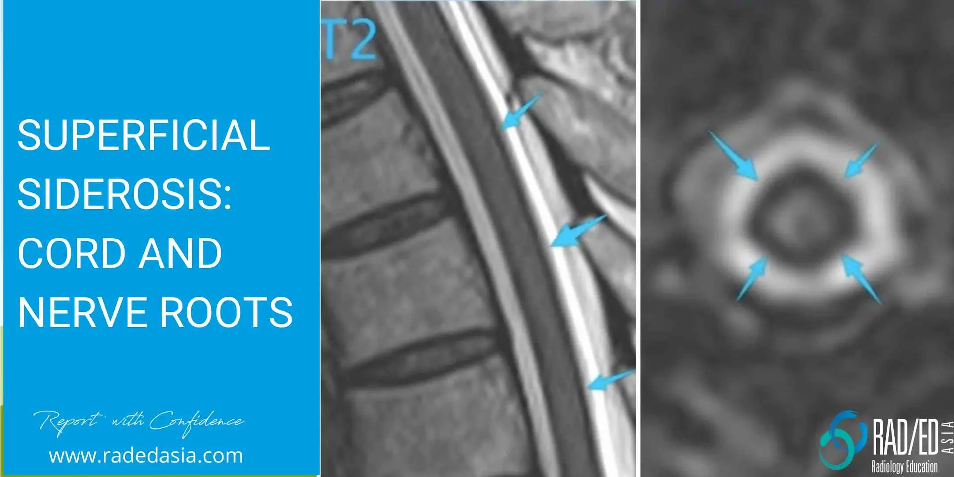
Superficial siderosis is the chronic deposition of hemosiderin in the subpial layer of the brain and spine
- It's due to chronic, low grade bleeding and only uncommonly due to an acute subarachnoid haemorrhage.
- The cause can be difficult to determine but often patients have had trauma or trans-dural surgery with resultant chronic low grade bleeding from those sites.
- Vascular malformations and rarely patients on long term warfarin with poor control can result in superficial siderosis.
- There is also an association with spinal epidural cysts which are thought to result from a tear in the dura resulting in low grade chronic bleeding and formation of an extra dural arachnoid cyst.
- Hemosiderin is low signal on T2 and Gradient Echo type sequences.
- Look for a rim of low signal outlining the cord (Blue and Pink arrows below).
Â
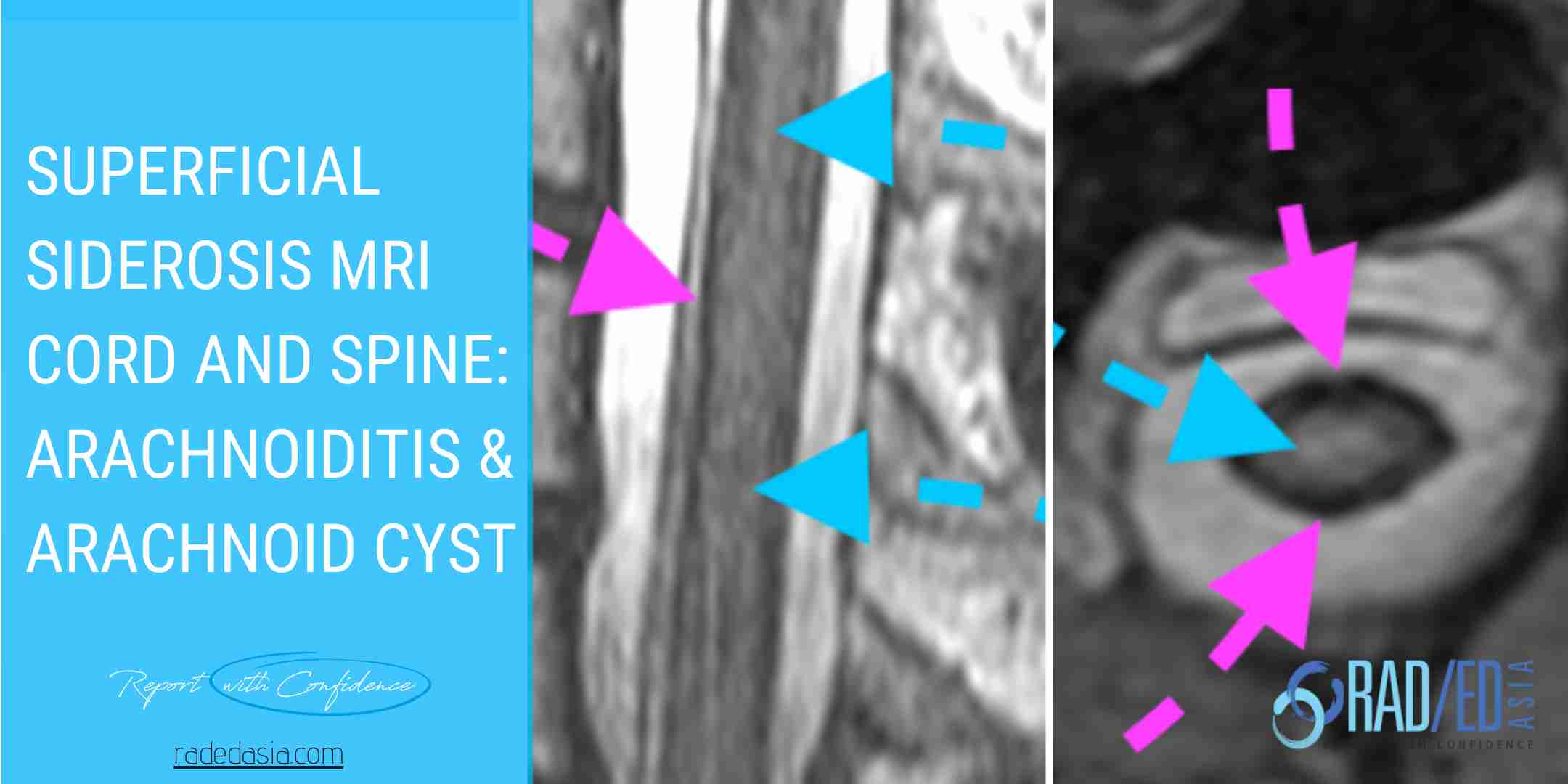
Hemosiderin deposition on Nerve Roots can result in arachnoiditis. Look for:
- Low signal of nerve roots (Orange arrow).
- Clumping and abnormal distribution of nerve roots (Green Arrow and centre image with asymmetric nerve root distribution).
- Enhancement of nerve roots.
Â
- If you see hemosiderosis on a spinal MRI look for an underlying cause.
- You need to image the whole spine and also the brain to look for a cause of the chronic bleeding.
We look at the complications of Superficial Siderosis in the next post.
Learn more about this condition & how best to report it in more detail in our SPINE & SIJ Imaging Mini Fellowships.
Click on the image below for more information.
- Join our WhatsApp RadEdAsia community for regular educational posts at this link: https://bit.ly/radedasiacommunityÂ
- Get our weekly email with all our educational posts: https://bit.ly/whathappendthisweek