This site is intended for Medical Professions only. Use of this site is governed by our Terms of Service and Privacy Statement which can be found by clicking on the links. Please accept before proceeding to the website.

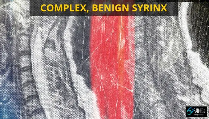
1. Hetrogeneous T2 signal: A large syrinx can have a heterogeneous T2 signal which is due to flow artefact within the syrinx.
2. Septations: Again, with a larger benign syrinx there can be considerable septation within the syrinx. However, there shouldn’t be nodularity and the septations shouldn’t enhance.
3. Eccentric position in cord: A benign syrinx can be central in the cord or eccentric in position.
4. Increasing size: An increase in size over time doesn’t mean its malignant. As long as there are no signs of malignancy, an increase in size is not an indicator of malignancy.
As long as there is no other feature of malignancy, such as cord oedema, enhancement, nodularity/ mass, the above findings should not be of concern for malignancy.
A very important point is that on the first presentation, even if the syrinx looks completely benign, you must give contrast. Tumors such as hemangioblastomas can present as a benign appearing syrinx on non-contrast scans, but on contrast administration, will show a tiny nodular area of enhancement. Once you have shown it to be benign on the first scan, follow up scans don’t require contrast.
Learn more about this condition & How best to report it in more detail in our SPINE Imaging Mini Fellowships.
Click on the image below for more information.

Stay tuned on new
Mini-Fellowships launches and learnings
This site is intended for Medical Professions only. Use of this site is governed by our Terms of Service and Privacy Statement which can be found by clicking on the links. Please accept before proceeding to the website.