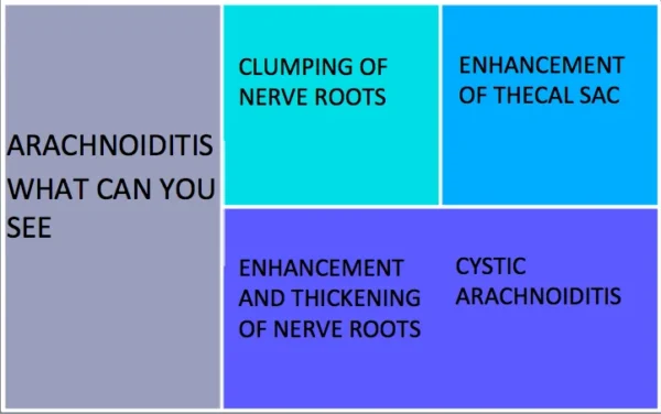

ARACHNOIDITIS ON MRI
What are some of the common appearances of arachnoiditis on MRI?
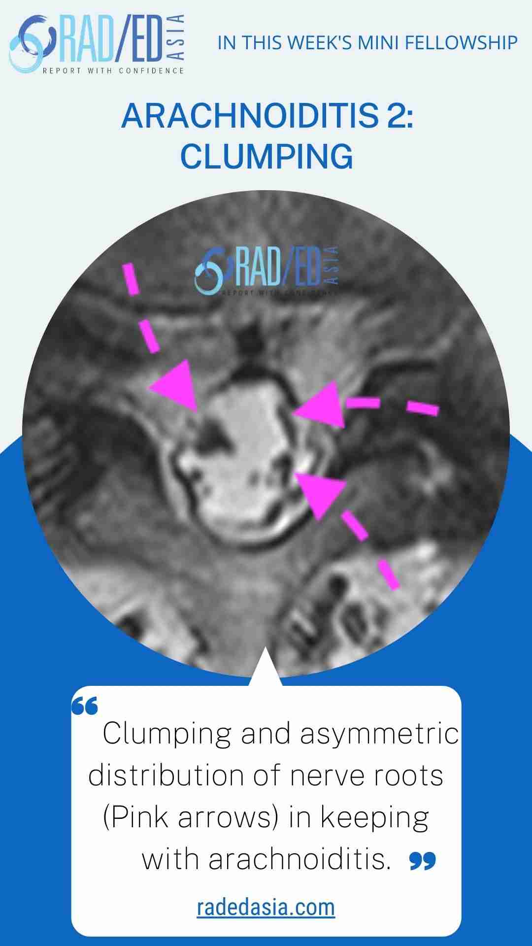
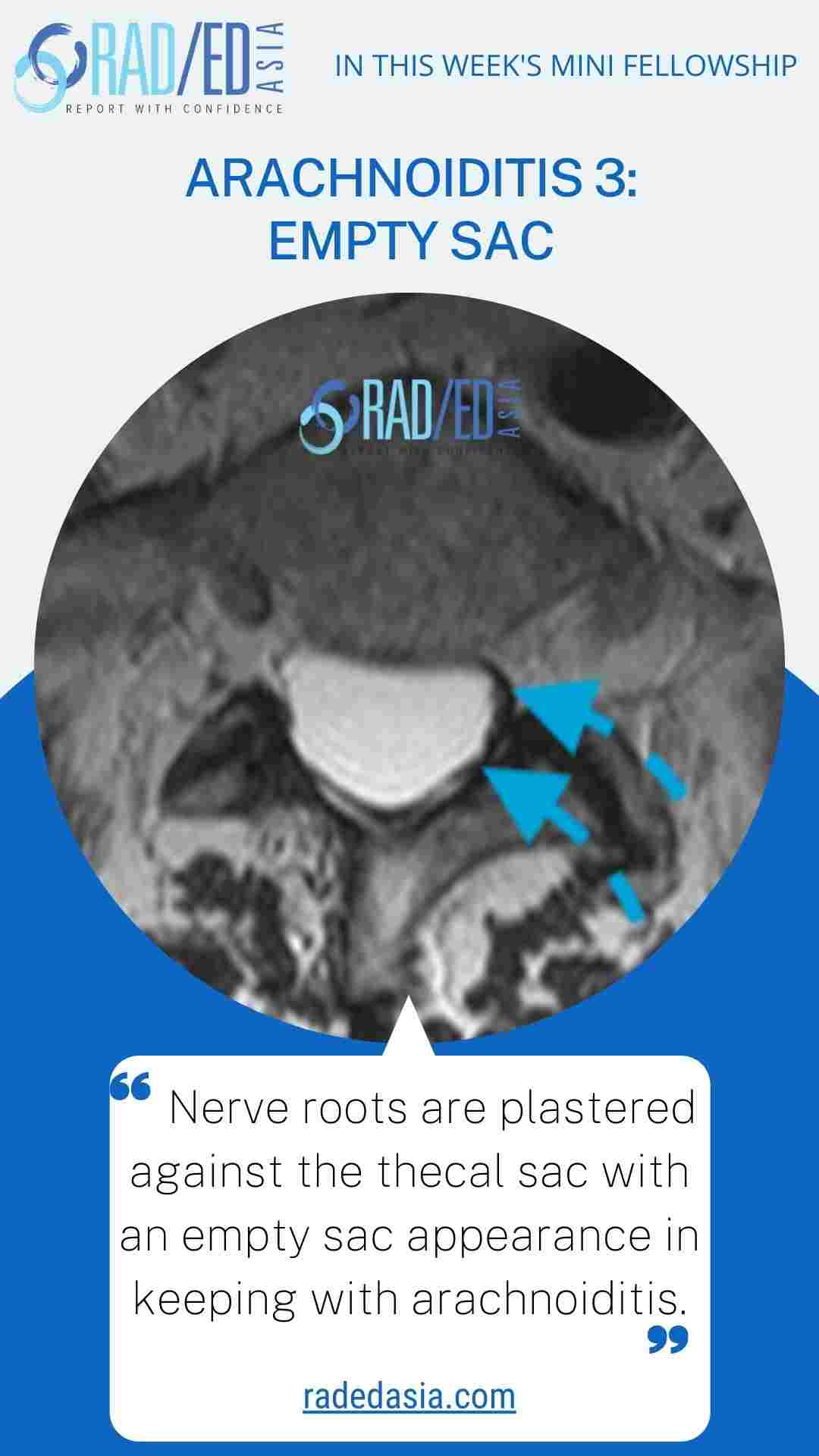
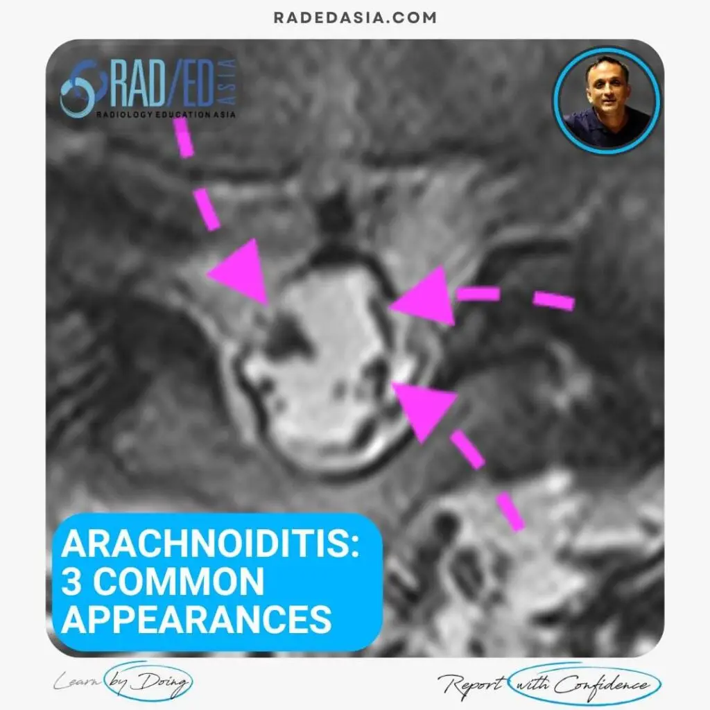
Another three quick images with some basic key points from our online spine MRI courses.
The distribution of nerve roots in the lumbar canal has to be symmetric. Any asymmetry indicates an abnormality.
Clumping in arachnoiditis occurs because fibrin lines the nerves post infection/ trauma/ bleeding which enables them to stick to each other.
The sac only appears empty as all the nerve roots are attached to the wall of the thecal sac.