CT scan of the cervical spine showing uncovertebral joint degeneration with sclerosis and loss of joint space coronal reformat

Stay tuned on new Fellowships and learnings

CT UNCOVERTEBRAL JOINT DEGENERATION CERVICAL SPINE

FROM TODAY'S REPORTING

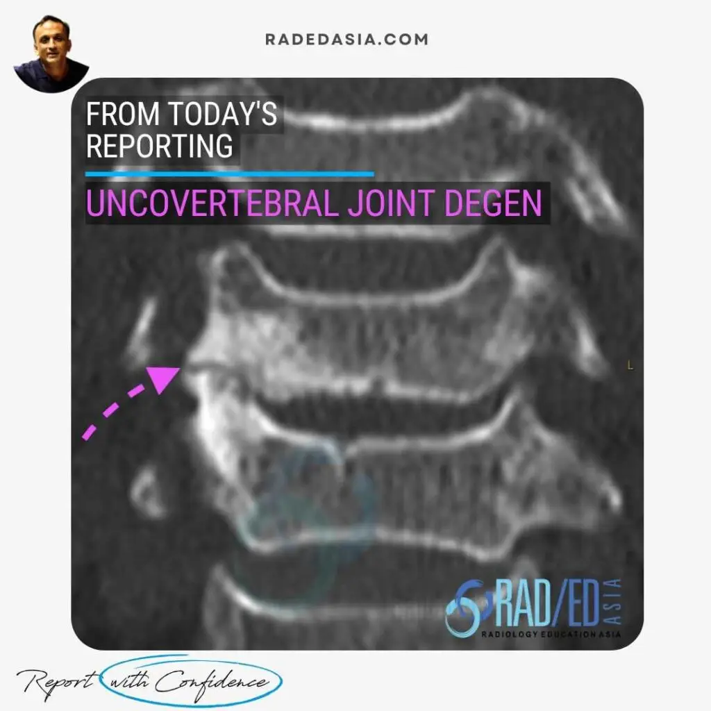
CERVICAL UNCOVERTEBRAL JOINT DEGENERATION
CORONAL CT


Demonstration reduction in joint space and significant subcortical sclerosis (Pink arrow) in the uncovertebral joint. Compare with normal contralateral uncovertebral joint where joint space is preserved and no sclerosis.


Uncovertebral joints best appreciated on coronal scans but osteophytes arising from them and narrowing the foramen best seen on sagittal scans.

We look at all of these topics in more detail in our Guided SPINE & SIJ Imaging Mini Fellowships.

Click on the image below for more information.

Our CPD & Learning Partners

OTHER POPULAR SPINE REPORTING POSTS: CLICK ON THE IMAGES BELOW

CT scan showing costotransverse joint degeneration with loss of joint space, subcortical sclerosis and cortical irregularity
PAIN MANAGEMENT
Axial CT image of the lumbar spine at the L4-L5 level showing focal calcification of the ligamentum flavum. Note both soft tissue thickening of the ligament flavum and the more dense calcification both contributing to right sided subarticular/lateral recess stenosis.
PAIN MANAGEMENT

Stay tuned on new Fellowships and learnings

error: Content is protected !!

Join our Newsletter

Stay tuned on new
Mini-Fellowships launches and learnings

Mini-Fellowships

Better Than Conferences

Join our WhatsApp Community

We help radiologists learn
by doing and report with confidence

This site is intended for Medical Professions only. Use of this site is governed by our Terms of Service and Privacy Statement which can be found by clicking on the links. Please accept before proceeding to the website.