SPINE DEGENERATION

SPINE DEGENERATION Learn Onlinelearn spine mri workshop online

All our posts on Spine & SIJ Degeneration Radiology . We regularly post on Spine MRI with all the posts on this page.

Spine Degeneration Radiology can be difficult due to the complex anatomy and the many pathologies that occur. In our posts we have concise explanation of the Spine MRI anatomy, the macroscopic pathology that’s relevant to the Imaging and the Imaging findings in various pathologies.

Learn where to Look, What to Look for and How to Report More Confidently

spine facet joint mri online radiology courses

SPINE MRI NERVE ROOT COMPRESSION ONLINE RADIOLOGY COURSE

KEY POINTS FROM OUR SPINE MRI COURSES Spine MRI Nerve Root Compression Online Radiology Course Another three quick images with some basic key points from our online spine mri courses. Here we look at how to evaluate and describe a disc that extends into the foramen and its relationship to the exiting foraminal nerve looking …

SPINE MRI NERVE ROOT COMPRESSION ONLINE RADIOLOGY COURSE Leer más »

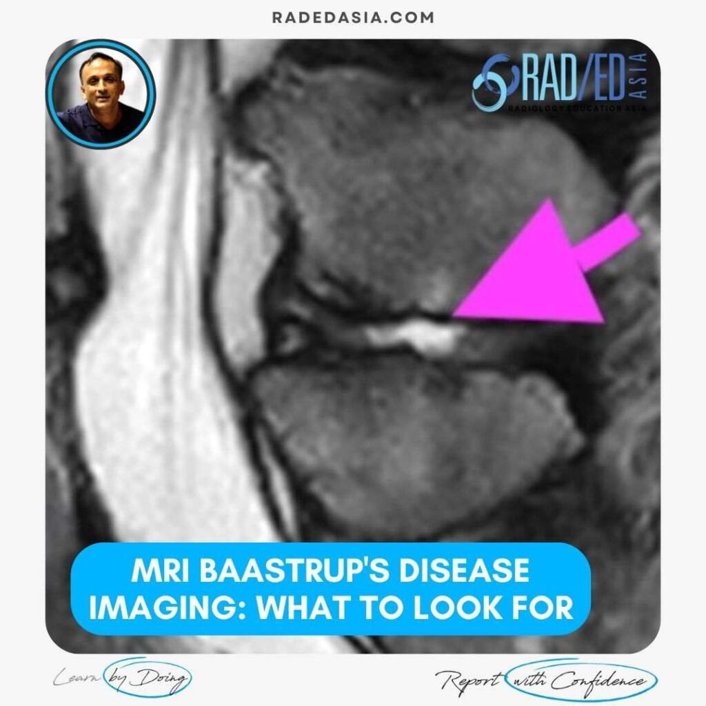
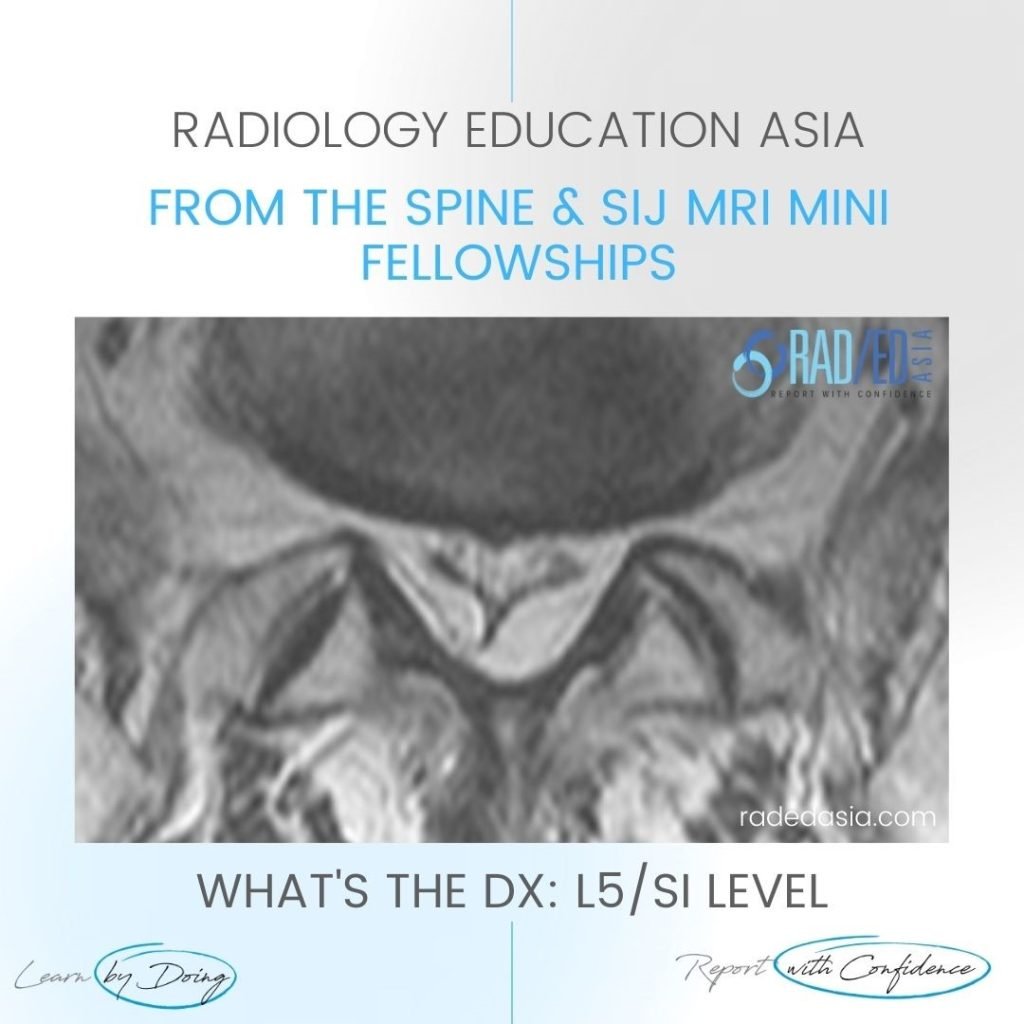
mri-baastrup's-disease-imaging-findings-what-to-look-for-radedasia

MRI BAASTRUP’S DISEASE: WHAT TO LOOK FOR

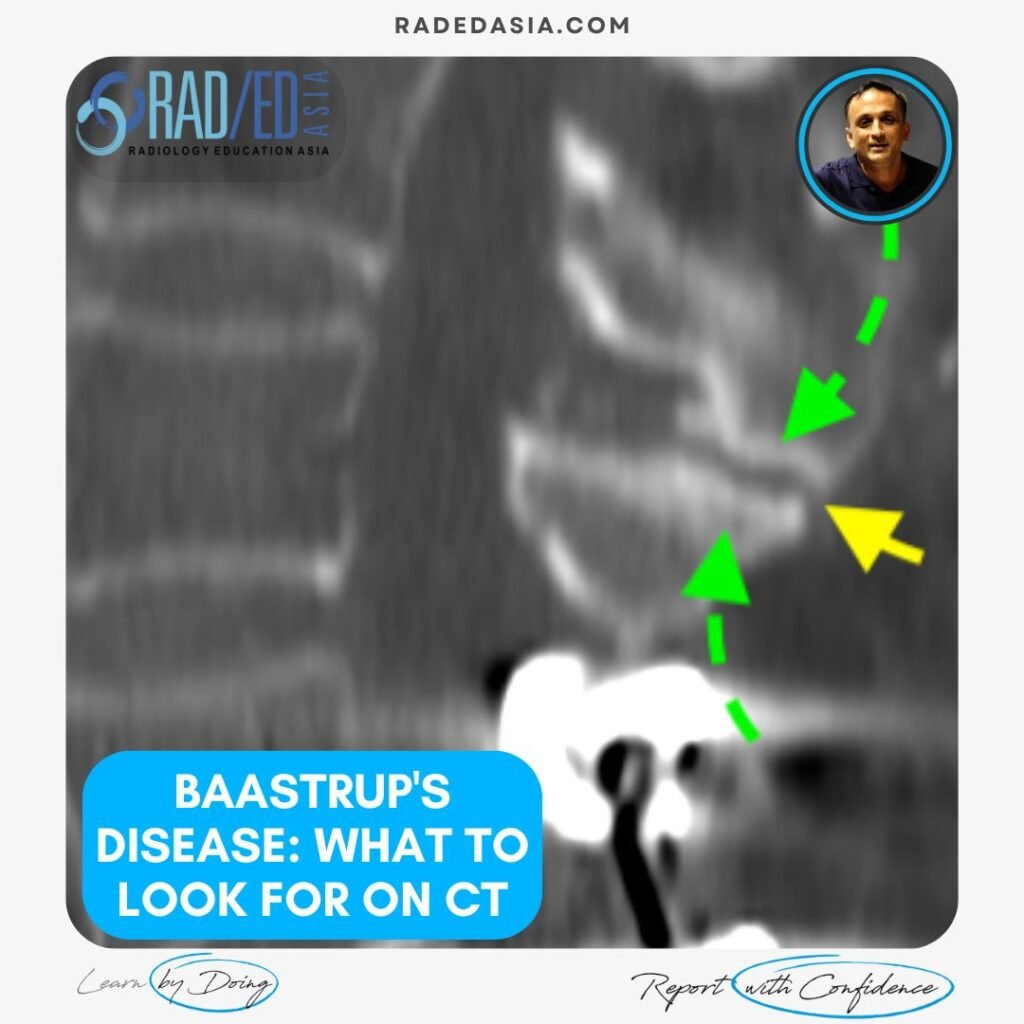
BAASTRUP'S DISEASE: WHAT TO LOOK FOR ON MRI Baastrup’s disease is caused by the friction of two spinous processes rubbing against each other and can result in central back pain but its difficult to diagnose without imaging. In the previous post we looked at the CT Findings in Baastrup’s Disease and this post looks at …

MRI BAASTRUP’S DISEASE: WHAT TO LOOK FOR Leer más »

spinal epidural lipomatosis mri lumbar thoracic radedasia

SPINAL EPIDURAL LIPOMATOSIS LUMBAR & THORACIC MRI

SPINAL EPIDURAL LIPOMATOSIS Spinal epidural lipomatosis is an excessive accumulation of non encapsulated fat in the epidural space. It can be seen on MRI in the thoracic or lumbar spine. In the cervical spine epidural lipomatosis is not seen. Epidural lipomatosis is not uncommon to see and is an important diagnosis to make as it …

SPINAL EPIDURAL LIPOMATOSIS LUMBAR & THORACIC MRI Leer más »

spine-thoracic-epidural-lipomatosis- radiology-spinal-spine-mri-radedasia

EPIDURAL SPACE INVADERS: SPINE MRI EPIDURAL LIPOMATOSIS PART 2

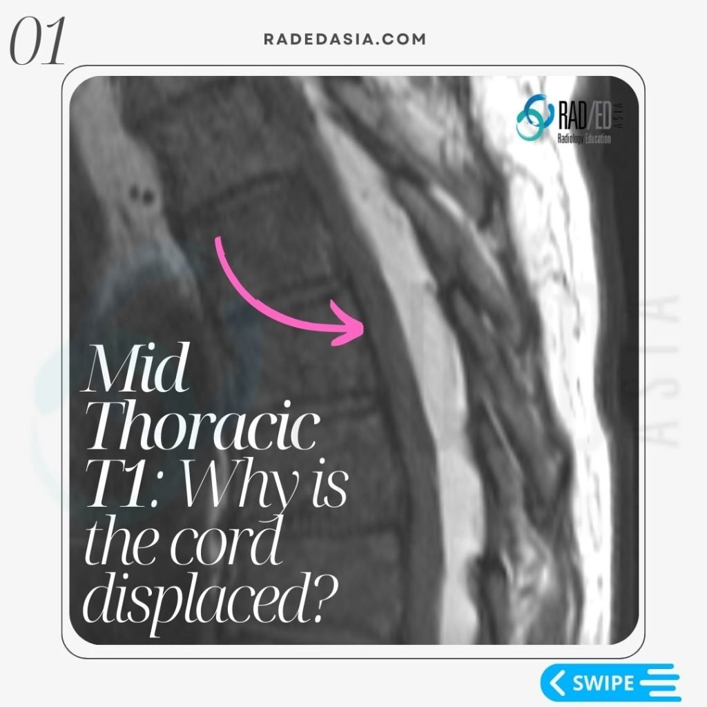
THORACIC EPIDURAL LIPOMATOSIS RADIOLOGY OVERVIEW Thoracic Epidural lipomatosis is not uncommon to see but has a different appearance to Lumbar Epidural Lipomatosis WHAT IS IT? With thoracic epidural lipomatosis there is excess fat accumulated in the epidural space. WHAT ARE THE CAUSES? There is an increased risk of developing epidural lipomatosis with: Chronic corticosteroid use. …

EPIDURAL SPACE INVADERS: SPINE MRI EPIDURAL LIPOMATOSIS PART 2 Leer más »

mri ct osteitis condensans ilii spine sij

OSTEITIS CONDENSANS ILII RADIOLOGY CT SACROILIAC JOINT SIJ (VIDEO)

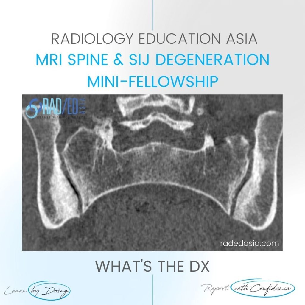
OSTEITIS CONDENSANS ILII RADIOLOGY CT SACROILIAC JOINT SIJ DISCUSSION ON OCI Osteitis Condensans Ilii.   Typical CT sclerotic changes of Osteitis Condensans Ilii. The sclerosis is continuous with the SIJ margin and particularly on the patient’s right it has a triangular appearance.   There is also mild sclerosis in the right sacrum adjacent to the …

OSTEITIS CONDENSANS ILII RADIOLOGY CT SACROILIAC JOINT SIJ (VIDEO) Leer más »

spine mri msk online radiology courses radedasia

LUMBAR SPINE ENHANCEMENT MRI ONLINE RADIOLOGY COURSES: KEY POINTS FROM OUR SPINE MRI COURSES

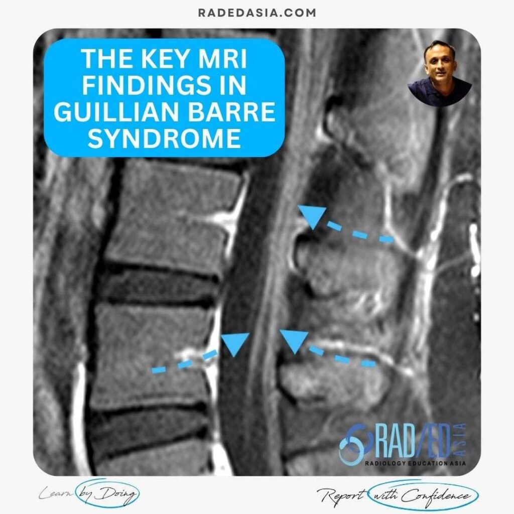
KEY POINTS FROM OUR SPINE MRI COURSES Spine MRI Online Radiology Course: Normal Spinal Enhancement Another three quick images with some basic key points from our online spine MRI courses. Here we look at areas of normal enhancement that can be mistaken for pathology: Normal enhancement of the anterior epidural plexus which can be mistaken …

LUMBAR SPINE ENHANCEMENT MRI ONLINE RADIOLOGY COURSES: KEY POINTS FROM OUR SPINE MRI COURSES Leer más »

opll spine ossification posterior longitudinal ligament ct mri xray radiology (1)

OPLL SPINE OSSIFICATION POSTERIOR LONGITUDINAL LIGAMENT CT (VIDEO)

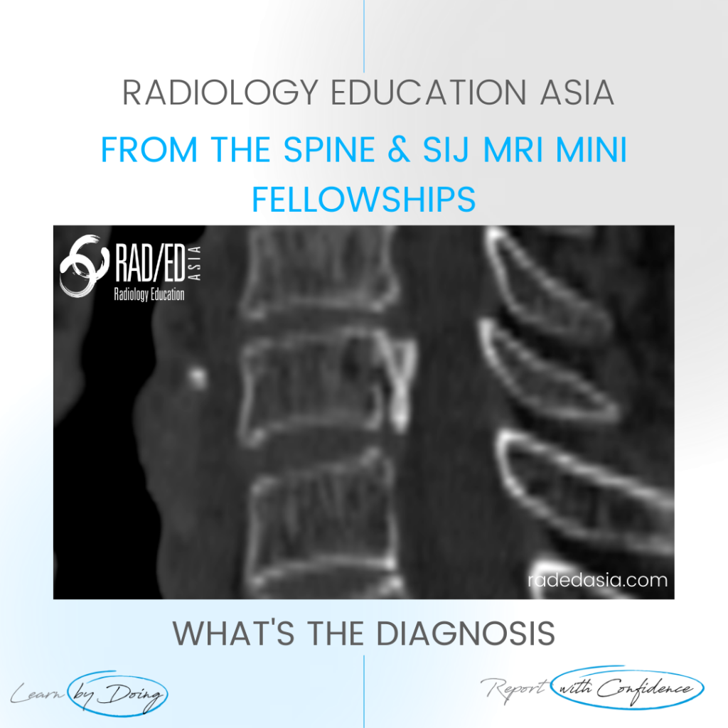
OPLL SPINE OSSIFICATION POSTERIOR LONGITUDINAL LIGAMENT CT DISCUSSION ON OPLL SPINE CT There is localised, linear ossification present at the posterior margin of the C6 vertebra.   This has a typical appearance of OPLL (Ossification of the Posterior Longitudinal Ligament).   The cervical spine is the most common location.   It is important to follow …

OPLL SPINE OSSIFICATION POSTERIOR LONGITUDINAL LIGAMENT CT (VIDEO) Leer más »

Este sitio está destinado únicamente a profesionales médicos. El uso de este sitio se rige por nuestra Términos de servicio y Declaracion de privacidad que se puede encontrar haciendo clic en los enlaces. Acepte antes de continuar con el sitio web.
error: Clic derecho no disponible.