meniscus tear mri vertical knee radedasia

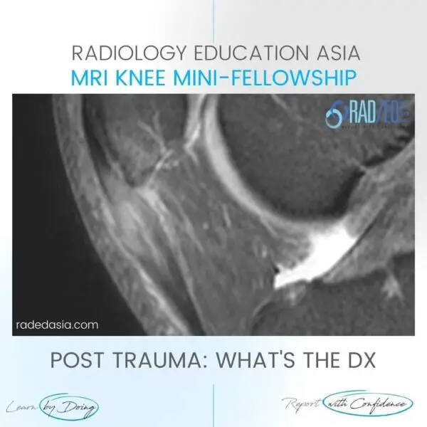
VERTICAL (LONGITUDINAL) MENISCUS TEAR POSTERIOR HORN.

  • The high signal extends to both the superior and inferior articular margins of the meniscus indicating a tear.


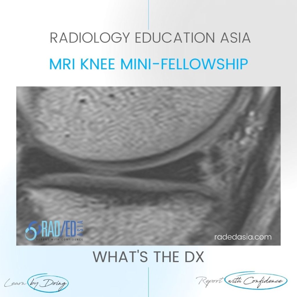
  • It can be easy to overlook peripheral vertical tears as the torn outer fragment (Green arrows) can be very thin and may not be interpreted correctly as torn meniscus.


  • Look for tissue that is the same signal as the parent meniscus and also irregularity of the residual meniscus margin which is torn.

Linear high signal extending from the inferior to superior articular surface (Pink arrows) of the posterior horn medial meniscus.

meniscus tear mri vertical knee radedasiaknee meniscus tear mri vertical radedasia

#meniscus #meniscustear #radiology #radedasia #mri #kneemri #msk #mriknee #mskmri #radiologyeducation #radiologycases #radiologist #radiologycme #radiologycpd #medicalimaging #imaging #radcme #rheumatology #arthritis #rheumatologist #sportsmed #orthopaedic #physio #physiotherapist

#radedasia #mri #mskmri #radiology

This site is intended for Medical Professionals only. Use of this site is governed by our Terms of Service and Privacy Statement which can be found by clicking on the links. Please accept before proceeding to the website.
error: Right Click not available.