This site is intended for Medical Professions only. Use of this site is governed by our Terms of Service and Privacy Statement which can be found by clicking on the links. Please accept before proceeding to the website.


MRI Capsulo Synovitis IGHL. At our MRI workshops we have an Open Mic policy. People are encouraged to interrupt and ask questions at any time because its best to clear doubts at the time topics are being discussed rather than wait till the end or never get to asking it at all.
One of the more consistent questions is about synovitis in the shoulder where certain sites are specifically involved but don’t have the typical appearance of synovitis we discussed in a previous post The Many Faces of Synovitis.
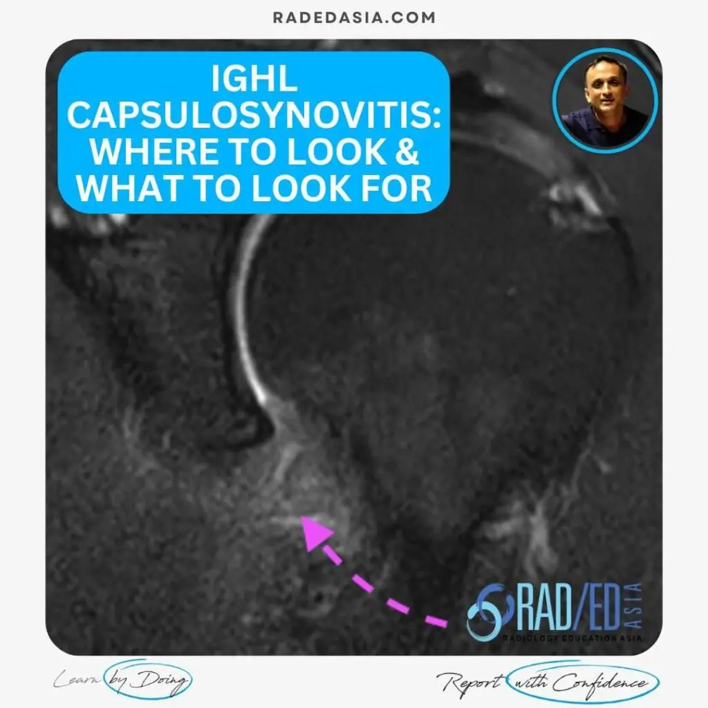
So, on MRI where do you look and what do you look for in Capsulo Synovitis in the Shoulder. In the shoulder there are two specific areas affected which are really a mixture of synovitis and capsulitis:

With capsulitis/synovitis, the IGHL thickens and becomes hyper-intense on the PD and PDFS scans.


Stay tuned on new
Mini-Fellowships launches and learnings
This site is intended for Medical Professions only. Use of this site is governed by our Terms of Service and Privacy Statement which can be found by clicking on the links. Please accept before proceeding to the website.