ANKLE
MRI OF PERIOSTEAL STRIPPING: INTRODUCTION
When evaluating ligament or tendon injuries on MRI, it’s easy to stop at identifying the tear. But one extra finding to look for is periosteal stripping — that can be missed if you’re not specifically assessing for it.
This post and video go through the MRI appearance, anatomical reasoning, and key patterns of periosteal stripping using practical trauma imaging examples, especially from the ankle and shoulder.
Read this post in conjunction with watching the video.
![]()
Periosteal stripping occurs when the periosteum is lifted off the cortex due to trauma. This happens at the attachment points of ligaments or tendons. Instead of the tendon or ligament rupturing it may remain intact but instead pulls the periosteum away from the underlying cortex.
Understanding the anatomy helps: the periosteum is normally tightly adhered to the cortex, forming a single, uninterrupted layer. When it gets stripped, this interface separates, and fluid or hemorrhage may accumulate in between, which becomes visible on MRI.
![]()
There are two key features to identify periosteal stripping on MRI:
1. Double Black Line Sign
On sequences like PD or T2 fat-saturated images, periosteal stripping creates two black lines:
Between these two, you may see high-signal fluid or edema.
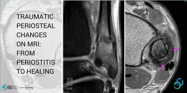
2. Edema at the Bone Margin
In the acute phase, edema and hemorrhage at the bone surface make the periosteal separation visible. Without edema, the periosteum and cortex blend together as a single dark structure, making it harder to detect.
If you spot posterior tibial margin edema in a case of syndesmotic injury, look closely — it often indicates periosteal lifting.
![]()
Case Example: Syndesmotic Injury and PITFL Tear
In a case of a posterior inferior tibiofibular ligament (PITFL) tear, the ligament may still appear continuous. However, rather than detaching directly from the bone, it pulls up the periosteum. On MRI, you’ll see:
![]()
Periosteal stripping can occur at any tendon or ligament attachment site and is common in the shoulder, especially in labral injuries, where similar patterns appear:
Recognizing this allows for more accurate grading of shoulder instability and labral avulsions.
![]()
Always look for periosteal stripping with ligament and tendon trauma.
When you identify periosteal stripping, your report should include:
This extra detail helps referring clinicians understand the true severity of injury.
![]()
![]()
Let’s start and we’re to look at ligament tears and periosteal stripping. Now ligament tears, I’m not going to go into the actual ligament tears themselves. This is all about you found a ligament tear, what more do you need to look at? And one of the things that you do need to look for is periosteal stripping and this is a pattern that we will see in a number of joints.
We’ve got a patient here who’s got a syndesmotic injury that’s a tear of the PITFL. What more do you need to look at? You can say in your report, can say it’s a torn PITFL. The referrer is probably going to be happy with it. But the thing is you need to do more. So the thing that happens with ligament attachments and also tendon attachments is that they attach to periosteum bone. So, sometimes instead of rupturing, what happens is that the periosteum itself gets lifted off the cortex. So that’s called periosteal stripping. And the way to understand that is if you look at this image here, so the dark yellow here is cortex, the lighter yellow is the periosteum. And there’s no plane in between them. They’re actually just of stuck together. And, this is a really lovely image because
this shows you is that this is the periosteum that’s been lifted off. They’ve cut it and lifted off and reflected it. And this is cortex underneath. But when we scan and we see periosteal stripping, this is what we see. We see a lifted off periosteum & we see cortex underneath it. And then the signal that we see, reflects this. So the normal periosteum edema, you can’t see it because the signal is exactly the same as cortex. So both are black on, this is a PDF headset, this is PD. So there’s a combination of periosteum and cortex here.
When you have periosteal stripping, there are two things that occur. One is because there’s trauma to the periosteum, you will get edema and hemorrhage. And when you see this pattern on the posterior margin, so someone has got a syndesmotic injury, if you see this pattern of edema and fluid tracking along the posterior margin of cortex almost for sure about periosteal stripping, this is what it does. It gives you this, track of fluid that goes along the back of the cortex or back of the tibia. The second thing to look at is to look at the actual periosteum. So now because the periosteum is not stuck to bone anymore, it’s been lifted off, you can now see it. So what we end up seeing is that we see two black lines. So here’s the cortical line and this really thin line here, that’s the strip periosteum. So now if I go back to this, This is what you’re seeing. So you’re seeing the thick black line of the cortex here and the thin black line of the lifted off periosteum here. Let’s go back to that MR. So thick cortex, thin periosteum.
What does that look like? So if we look at a scan here, so we’ve got a sagittal, I think this is a T2 fat sat. And as we scroll, what we end up seeing is that we’ve got a black line here, which is the then we have a second black line. Whenever in trauma, if you see two black lines, that means that it’s periosteal stripping and we also have edema as well.
This is really the key, when you see two black lines like this. And this is the space now that’s being created between the periosteum and the cortex that’s being lifted off.
If we look at this on the axial, so this patient’s had significant trauma, syndesmotic, sorry, syndesmotic injury. is completely torn. So it’s very severe trauma. But what we’re looking at here is actually the periosteum. So this is the periosteum that’s been lifted off anteriorly. And here’s the periosteum that’s been lifted off posteriorly. So you can see that the PTFL is still actually continuous.
All it’s done is that, the PTFL should be inserting around here. So instead it’s basically lifted, it’s still attached to the periosteum, but it’s just lifted it off and stripped it off the attachment to cortex.
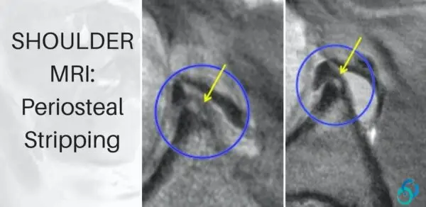
So now this is not the only place we see it. We also see it virtually everywhere else, but very commonly we see this in the shoulder. So this pattern again, so we’ve got a labrum here. Here’s a labrum then you see cortex and then you see a second black line. So this is the periosteum that’s been stripped. So the labrum is still attached to the periosteum. So this is periosteum, this is labrum and it’s stripped off the attachment and then fluid is then tracking between stripped off periosteum and the cortex, which is exactly the same appearance that we saw just before in the ankle.
Okay, so for periosteal stripping, the key points are, one, in a normal scan, you don’t see the periosteum. But in the context of trauma, if you see a double, look for a double black line. The first thing is actually look for edema. Look for edema at the bone margins, because if you don’t have edema, you’re not going to have periosteal stripping in the acute stage. Once you see the edema, then look for the double black line. That tells you that that’s stripping of the periosteum.
![]()