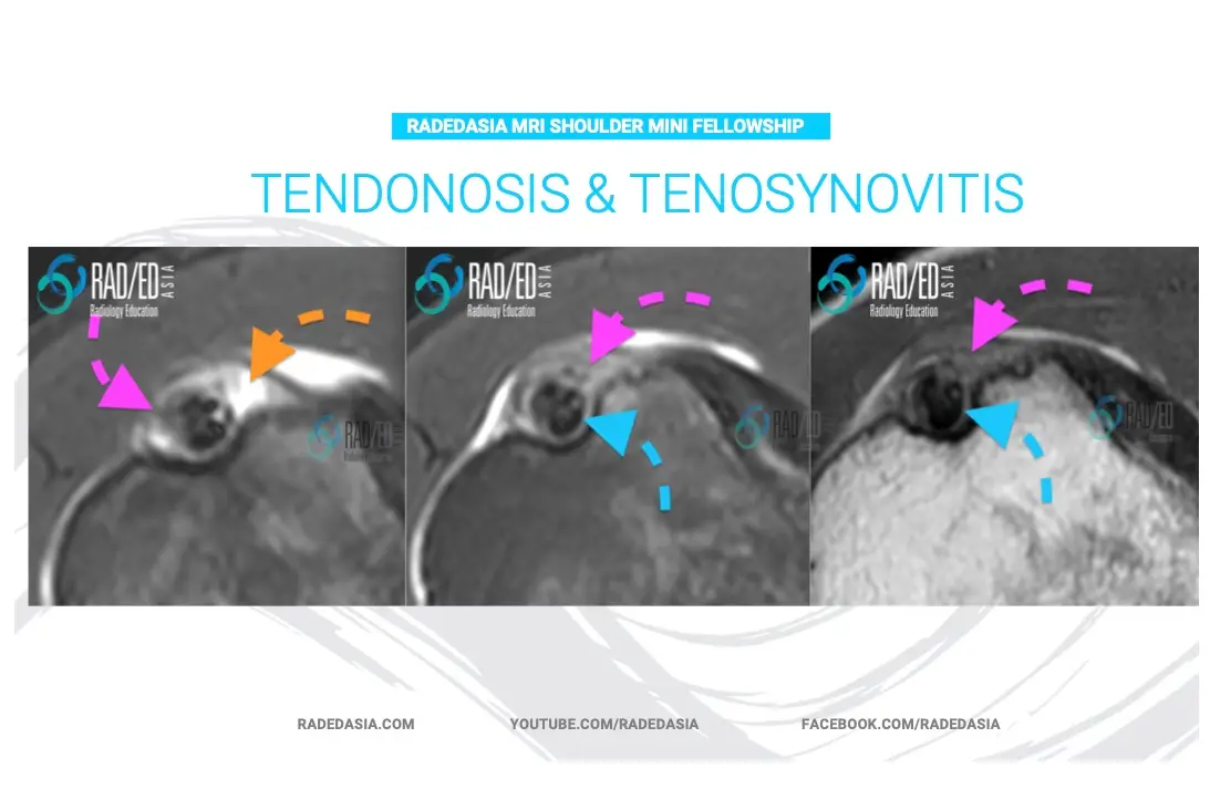
We are looking at MRI of Tendonosis / Tendonitis and Tenosynovitis of the Biceps Tendon this week in the MRI Shoulder Mini fellowship.Here is a pattern of changes in the Biceps tendon sheath but can be used in most tendons in the body. 1. Uncomplicated tendon sheath fluid is bright on PD/T2 and T2FS/ PDFS (Orange Arrow).2. Synovitis/ Synovial thickening becomes intermediate in signal (Pink Arrow).3. Tendonosis is Intermediate signal on PD/T2 (Blue arrow) and may or may not be associated with enlargement of the tendon.
This site is intended for Medical Professions only. Use of this site is governed by our Terms of Service and Privacy Statement which can be found by clicking on the links. Please accept before proceeding to the website.