SEO

mri ankle foot ligament short plantar

MRI PLANTAR LIGAMENT SHORT PLANTAR LIGAMENT ANKLE FOOT AND TOE (VIDEO)

MRI PLANTAR LIGAMENT SHORT PLANTAR LIGAMENT WHAT'S THE STRUCTURE The Short Plantar Ligament. WHAT ARE THE FINDINGS ANATOMY OF THE SHORT PLANTAR LIGAMENT. 1. Extends from the calcaneus (Green star) to the cuboid (Yellow star). 2. It is distal and separate to long plantar ligament. 3. Has a fan shaped appearance. MRI PLANTAR LIGAMENT SHORT […]

MRI PLANTAR LIGAMENT SHORT PLANTAR LIGAMENT ANKLE FOOT AND TOE (VIDEO) Read More »

subcortical subchondral fracture knee tibial plateau mri insufficiency

SUBCORTICAL SUBCHONDRAL FRACTURE KNEE MRI TIBIAL PLATEAU (VIDEO)

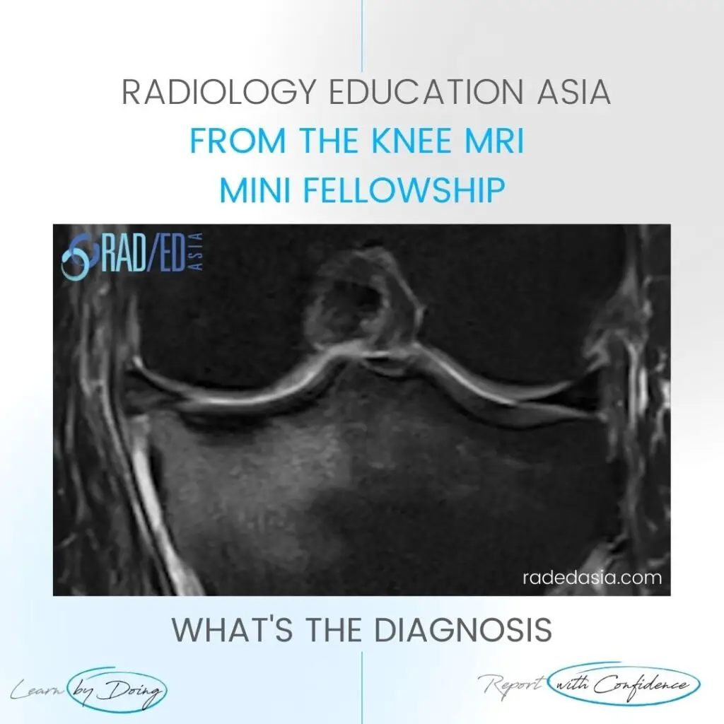
SUBCORTICAL SUBCHONDRAL FRACTURE KNEE MRI TIBIAL PLATEAU DISCUSSION Subchondral or subcortical fractures of the knee can occur in the femoral condyles or tibial plateau. The key to the diagnosis is seeing a linear low signal line adjacent to and paralleling the cortex without any cortical defect or break in the acute phase.   There is usually

SUBCORTICAL SUBCHONDRAL FRACTURE KNEE MRI TIBIAL PLATEAU (VIDEO) Read More »

mri ct osteitis condensans ilii spine sij

OSTEITIS CONDENSANS ILII RADIOLOGY CT SACROILIAC JOINT SIJ (VIDEO)

OSTEITIS CONDENSANS ILII RADIOLOGY CT SACROILIAC JOINT SIJ DISCUSSION ON OCI Osteitis Condensans Ilii. Typical CT sclerotic changes of Osteitis Condensans Ilii. The sclerosis is continuous with the SIJ margin and particularly on the patient’s right it has a triangular appearance. There is also mild sclerosis in the right sacrum adjacent to the SIJ (Green

OSTEITIS CONDENSANS ILII RADIOLOGY CT SACROILIAC JOINT SIJ (VIDEO) Read More »

shoulder labrum mri variant anatomy bicipito labrum complex radedasia

SHOULDER LABRUM MRI VARIANT ANATOMY BICIPITO LABRUM COMPLEX: Don’t call these three appearances of the labrum a tear.

SHOULDER MRI LABRUM MRI VARIANT ANATOMY There are a number of normal appearances of the Bicipito Labral Complex which can mistakenly be called tears.   Here is what they are and where to look for them.   (These posts are Part of our Quick Pick Series with concise information from the Journals I read). CLICK

SHOULDER LABRUM MRI VARIANT ANATOMY BICIPITO LABRUM COMPLEX: Don’t call these three appearances of the labrum a tear. Read More »

mri musculotendinous junction cysts and delaminating rotator cuff tears

MRI DELAMINATION ROTATOR CUFF TEARS MUSCULOTENDINOUS JUNCTION CYSTS

MRI DELAMINATING ROTATOR CUFF TEARS & MUSCULOTENDINOUS JUNCTION CYSTS On MRI Rotator cuff delaminating tears can result in Musculotendinous Junction Cysts.   These Musculotendinous junction cysts are seen in the rotator cuff tendons, mostly in Supraspinatus and less commonly in infraspinatus.   In this post we look at the appearance of both Delaminating Rotator cuff

MRI DELAMINATION ROTATOR CUFF TEARS MUSCULOTENDINOUS JUNCTION CYSTS Read More »

tibialis posterior tendinitis tendinopathy tear mri radiology

TIBIALIS POSTERIOR TENDINITIS TENDINOPATHY TEARS RADIOLOGY MRI (VIDEO)

TIBIALIS POSTERIOR TENDINITIS TENDINOPATHY TEARS RADIOLOGY MRI WHAT ARE THE FINDINGS The tibialis posterior tendon is enlarged and increased in signal. DIAGNOSIS & DISCUSSION TIBIALIS POSTERIOR TENDINOPATHY AND PARTIAL TEAR. Tibialis posterior tendinopathy and delamination / partial tears. The normal tibialis posterior tendon should be uniformly low signal. Enlargement and or increased intermediate signal indicates

TIBIALIS POSTERIOR TENDINITIS TENDINOPATHY TEARS RADIOLOGY MRI (VIDEO) Read More »

opll spine ossification posterior longitudinal ligament ct mri xray radiology (1)

OPLL SPINE OSSIFICATION POSTERIOR LONGITUDINAL LIGAMENT CT (VIDEO)

OPLL SPINE OSSIFICATION POSTERIOR LONGITUDINAL LIGAMENT CT DISCUSSION ON OPLL SPINE CT There is localised, linear ossification present at the posterior margin of the C6 vertebra. This has a typical appearance of OPLL (Ossification of the Posterior Longitudinal Ligament). The cervical spine is the most common location. It is important to follow this up with

OPLL SPINE OSSIFICATION POSTERIOR LONGITUDINAL LIGAMENT CT (VIDEO) Read More »

ischiofemoral impingement radiology mri hip

ISCHIOFEMORAL IMPINGEMENT RADIOLOGY HIP MRI (VIDEO)

ISCHIOFEMORAL IMPINGEMENT HIP MRI RADIOLOGY ISCHIOFEMORAL IMPINGEMENT: WHY DOES IT OCCUR Ischiofemoral impingement is a result of the quadratus femoris muscle being compressed between the ischial tuberosity and the posterior femur resulting initially in quadratus femoris oedema but can progress to tears and severe atrophy. ISCHIOFEMORAL IMPINGEMENT: WHAT ARE THE FINDINGS Initially there is oedema

ISCHIOFEMORAL IMPINGEMENT RADIOLOGY HIP MRI (VIDEO) Read More »

meniscus tear maceration degeneration mri knee radedasia

MENISCUS TEAR MACERATION DEGENERATION MRI KNEE RADIOLOGY (VIDEO)

MENISCUS MACERATION DEGENERATION MRI KNEE WHAT ARE THE FINDINGS MENISCUS MACERATION DEGENERATION MRI KNEE   Generalized increase in meniscal signal. Intermediate increase in signal on PD and PD/T2FS. The meniscus generally retains it’s shape but is ill defined. Due to the degeneration its more likely to tear and it also stops functioning like a normal

MENISCUS TEAR MACERATION DEGENERATION MRI KNEE RADIOLOGY (VIDEO) Read More »

annular fissure tear mri radiology lumbar spine radedasia

ANNULAR FISSURE TEAR MRI RADIOLOGY LUMBAR SPINE (VIDEO)

Stay tuned on new Fellowships and learnings Subscribe The Dx / Spine Degeneration ANNULAR FISSURE TEAR MRI RADIOLOGY HOW DO YOU FIND AN ANNULAR FISSURE ANNULAR FISSURE ON MRI Annular fissures on MRI are relatively common. Whilst they don’t result in radiculopathy, they can be a cause of back pain and are important to diagnose

ANNULAR FISSURE TEAR MRI RADIOLOGY LUMBAR SPINE (VIDEO) Read More »

error: Content is protected !!

Join our Newsletter

Stay tuned on new
Mini-Fellowships launches and learnings

Mini-Fellowships

Better Than Conferences

Join our WhatsApp Community

We help radiologists learn
by doing and report with confidence

This site is intended for Medical Professions only. Use of this site is governed by our Terms of Service and Privacy Statement which can be found by clicking on the links. Please accept before proceeding to the website.