subcortical subchondral fracture knee tibial plateau mri insufficiency
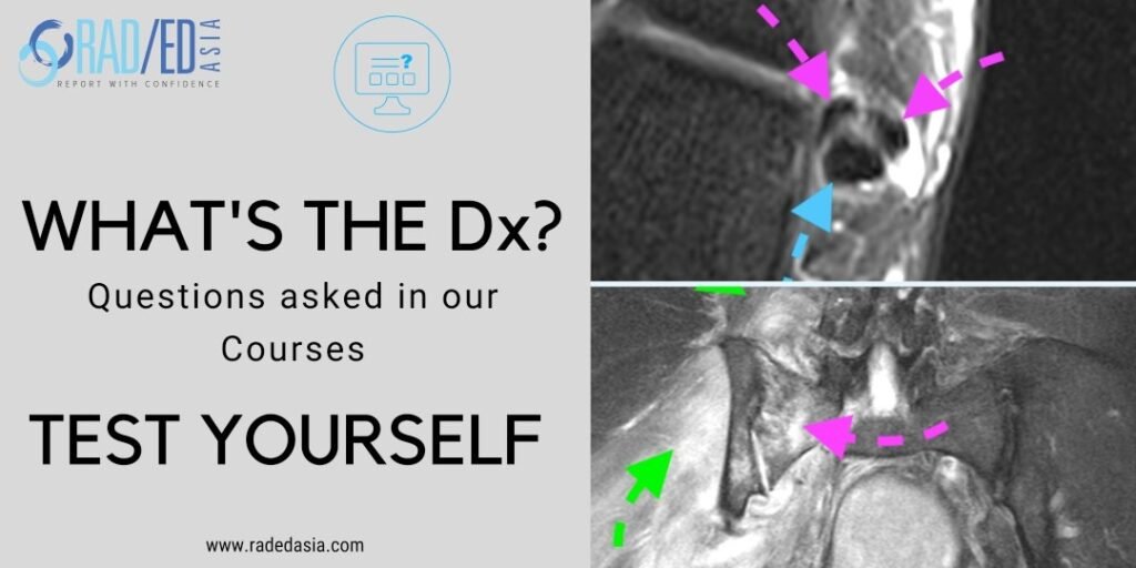
  • Subchondral or subcortical fractures of the knee can occur in the femoral condyles or tibial plateau.


  • The key to the diagnosis is seeing a linear low signal line adjacent to and paralleling the cortex without any cortical defect or break in the acute phase.  


  • There is usually significant bone marrow oedema spreading around the fracture line.


  • An older term for this appearance is SONK (Spontaneous Osteonecrosis of the Knee) however the pathology is now recognised to commence with a subcortical fracture.


  • More chronically osteonecrosis can occur in the region of the fracture with collapse of the cortex.
    • When this occurs there will be contour deformity of the cortex.

  • Extensive bone marrow oedema tibial plateau (Yellow arrows) and,


  • Linear low signal subcortical line which is low signal on T2FS and T1 (Green arrows) which is a subcortical fracture.


  • Additional and not directly related extrusion of the meniscus (Pink arrow).

Learn more about KNEE Imaging in our ONLINE or ONSITE
Guided MRI KNEE Mini-Fellowship.


More by clicking on the images below.

acl-pcl-mucoid-degeneration-mri-findings-radedasia
Anterior Cruciate Ligament
mri knee meniscus degeneration maceration flap tear extrusion meniscocapsular separation radedasia
KNEE

#radiology #radiologist #radiologia #mri #kneemri #mskmri #msk #mskrad #mskradiology #imaging #frcr #sportsmed #radiologyresident #foamrad #emergencydepartment #ortho #ct #radiologystudent #trauma #radedasia #radiologycme #radiologyeducation #radiologycases #rheumatology #arthritis #painphysician #chiropractic #physiotherap #kneesurgeon

#radedasia #mri #mskmri #radiology

This site is intended for Medical Professionals only. Use of this site is governed by our Terms of Service and Privacy Statement which can be found by clicking on the links. Please accept before proceeding to the website.
error: Right Click not available.