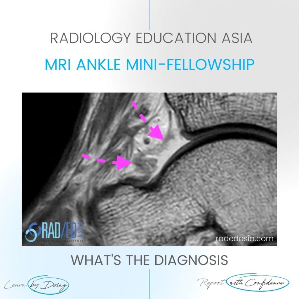
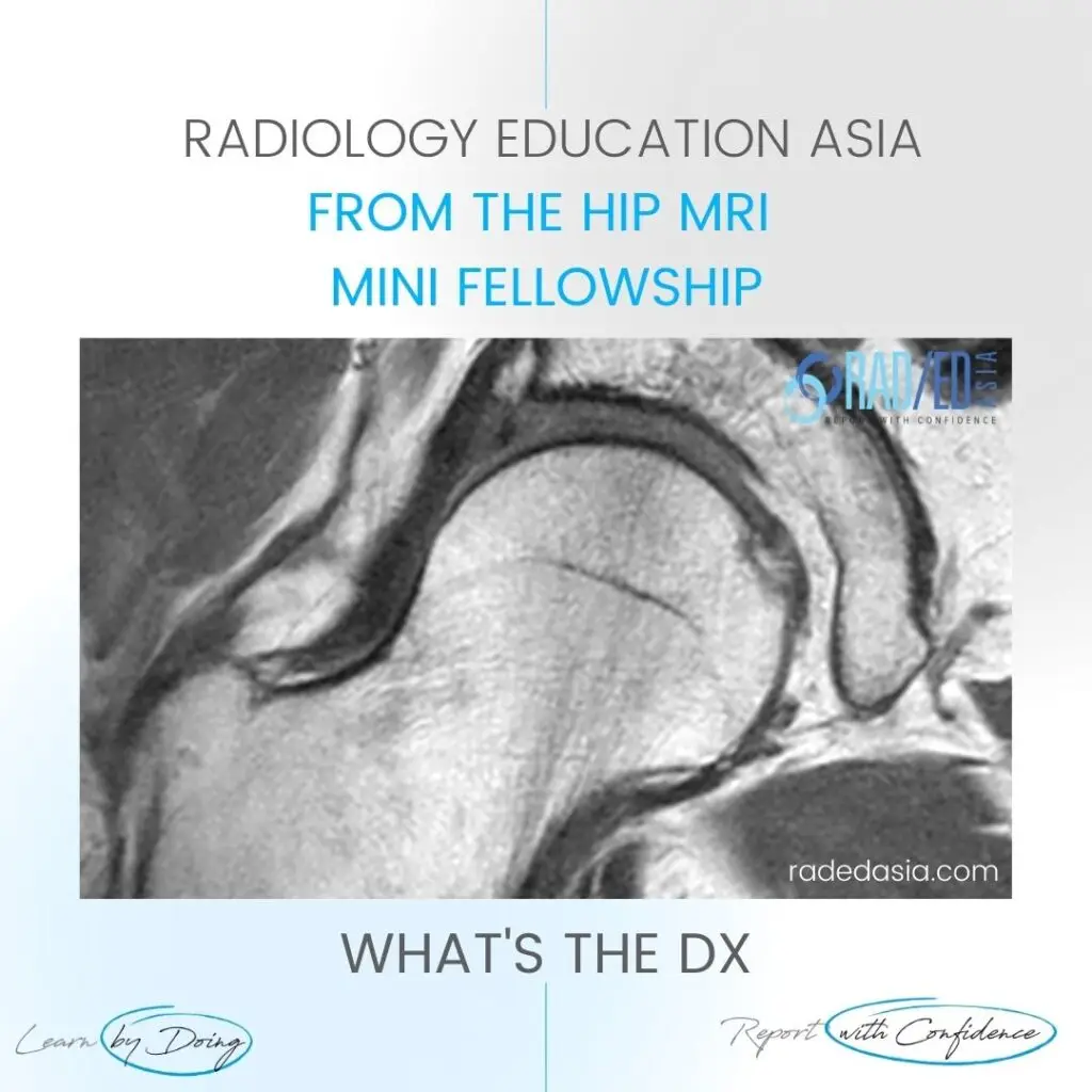
Dx

learn knee popliteomeniscal fasicles normal radedasia

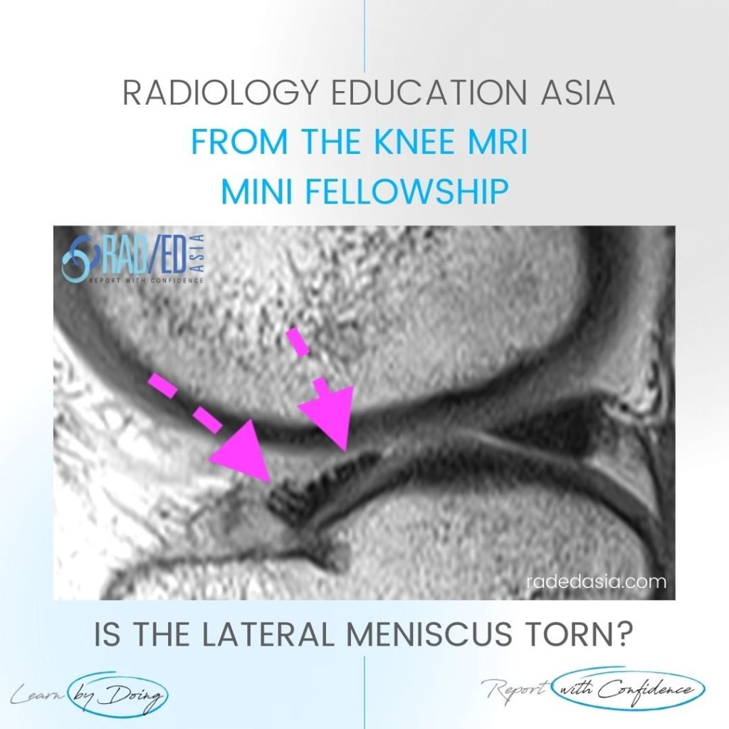
POPLITEOMENISCAL FASICLES MRI TEAR NORMAL ANATOMY

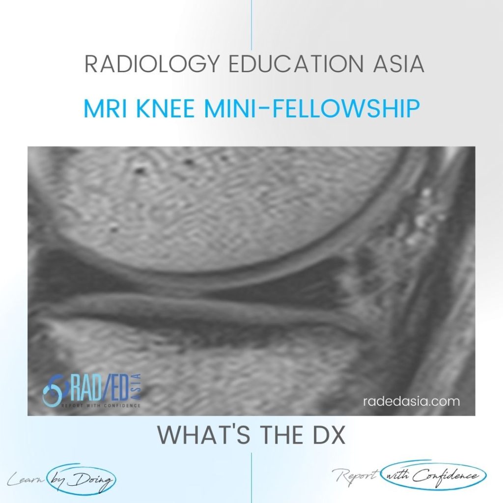
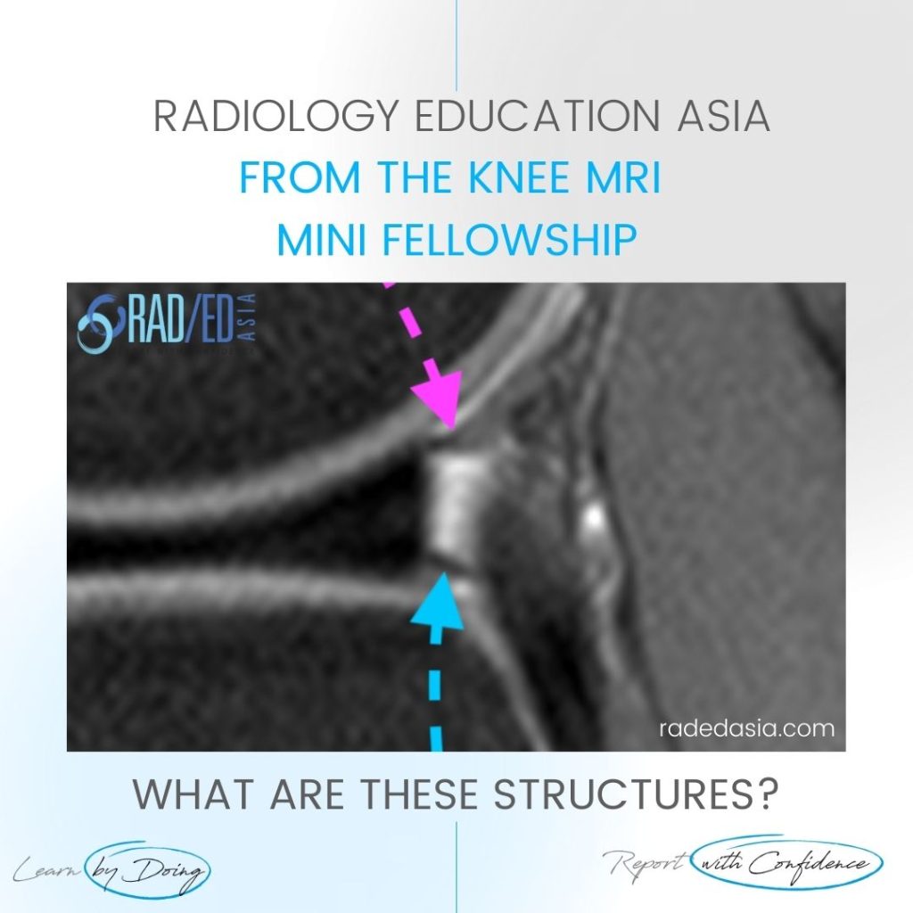
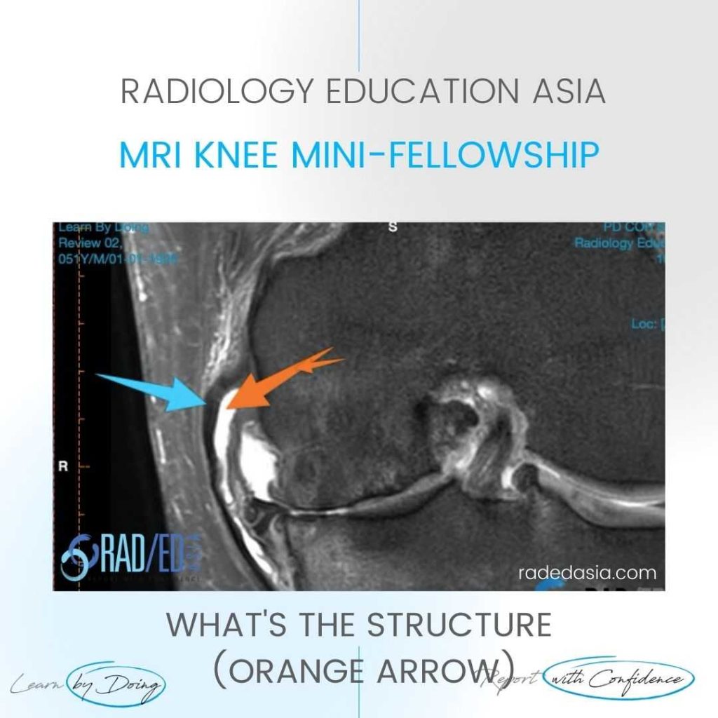
POPLITEOMENISCAL FASICLES MRI TEAR NORMAL ANATOMY DISCUSSION These structures are the Popliteomeniscal Fascicles.   They are the Antero Inferior Fascicle (Blue arrow).   The Postero superior Fascicle (Pink arrow) which forms the,   Popliteal Hiatus (Orange arrow) for the,   Popliteus tendon (Green arrow)  to pass through. POPLITEOMENISCAL FASICLES MRI TEAR NORMAL ANATOMY: MOVE SLIDER

POPLITEOMENISCAL FASICLES MRI TEAR NORMAL ANATOMY Read More »

shoulder labrum normal variant buford complex sublabral foramen hole mri

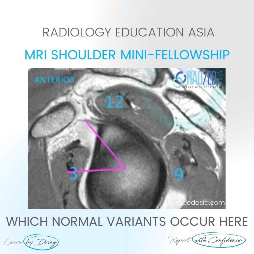
SHOULDER LABRUM NORMAL VARIANTS: WHICH VARIANTS OCCUR HERE (VIDEO)

SHOULDER LABRUM NORMAL VARIANTS DISCUSSION Sublabral Foramen & Buford Complex. FINDINGS Two normal variants occur in superior portion of the anterior labrum: Sublabral Foramen (also called Sublabral Hole), The Buford complex. SHOULDER LABRUM NORMAL VARIANTS VIDEO https://youtu.be/LDjtG2x_XEE If your Browser is blocking the video, Please view it on our YouTube Channel HERE. Learn more about

SHOULDER LABRUM NORMAL VARIANTS: WHICH VARIANTS OCCUR HERE (VIDEO) Read More »

medullary infarct radiology femur tibia mri radedasia

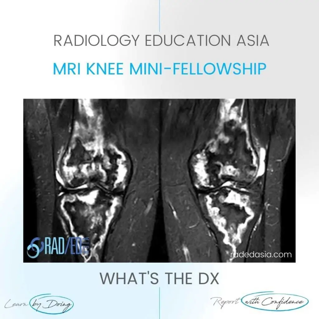
MEDULLARY BONE INFARCTS RADIOLOGY KNEE MRI

MEDULLARY BONE INFARCTS RADIOLOGY KNEE MRI DISCUSSION MEDULLARY BONE INFARCTS. This is a characteristic appearance of medullary bone infarcts. Most commonly they are in the diaphysis and metaphysis but in this case they also extend to the epiphysis.   The important think to note here is that its involving multiple areas. When there are multiple

MEDULLARY BONE INFARCTS RADIOLOGY KNEE MRI Read More »

learn shoulder mri biseps disloc cyst radedasia

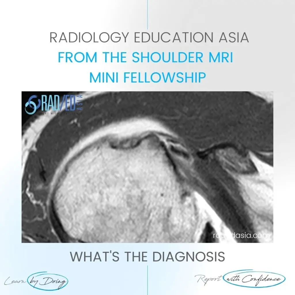
BICEPS DISLOCATION MRI SHOULDER RADIOLOGY

BICEPS DISLOCATION MRI SHOULDER RADIOLOGY DISCUSSION ON BICEPS DISLOCATION Extra articular Biceps dislocation. This is an Extra articular Biceps dislocation into a partial tear the Subscapularis tendon. FINDINGS IN BICEPS DISLOCATION Empty Bicipital Groove (Blue arrow).   Biceps tendon dislocated (Pink arrow).   Partial tear of Subscapularis tendon (Green arrow). High signal is seen within the

BICEPS DISLOCATION MRI SHOULDER RADIOLOGY Read More »

error: Content is protected !!

Join our Newsletter

Stay tuned on new
Mini-Fellowships launches and learnings

Mini-Fellowships

Better Than Conferences

Join our WhatsApp Community

We help radiologists learn
by doing and report with confidence

This site is intended for Medical Professions only. Use of this site is governed by our Terms of Service and Privacy Statement which can be found by clicking on the links. Please accept before proceeding to the website.