Dx

freiberg disease mri radiology toe radedasia

FREIBERG DISEASE SUBCHONDRAL FRACTURE INFRACTION OSTEOCHONDROSIS (VIDEO)

FREIBERG DISEASE / SUBCHONDRAL FRACTURE INFRACTION / OSTEOCHONDROSIS DISCUSSION FREIBERG DISEASE / SUBCHONDRAL FRACTURE INFRACTION / OSTEOCHONDROSIS Extensive oedema in the head of the 2nd metatarsal with slight flattening of the cortical margin is characteristic of what’s called Freiberg’s Disease in adolescents and Subchondral Fracture in adults. Freiberg disease occurs in adolescents with an unfused …

FREIBERG DISEASE SUBCHONDRAL FRACTURE INFRACTION OSTEOCHONDROSIS (VIDEO) Read More »

knee infrapatellar plica syndrome radiology mri radedasia

KNEE INFRAPATELLAR PLICA SYNDROME RADIOLOGY MRI (VIDEO)

KNEE INFRAPATELLAR PLICA SYNDROME RADIOLOGY MRI DISCUSSION ON KNEE INFRAPATELLAR PLICA PLICAS IN THE KNEE The infrapatellar plica is one of 4 plicas in the knee: Medial Patella Plica. Suprapatellar Plica. Infrapatellar plica. Lateral Patellar plica. INFRAPATELLAR PLICA ANATOMY Attaches to anterior intercondylar notch and inferior pole patella.   Runs through Hoffa’s Fat Pad.   …

KNEE INFRAPATELLAR PLICA SYNDROME RADIOLOGY MRI (VIDEO) Read More »

osgood schlatter xray radiology radedasia

OSGOOD SCHLATTER RADIOLOGY X-RAY (VIDEO)

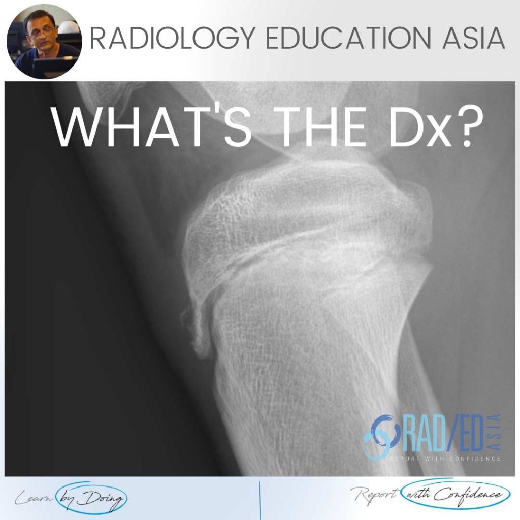
OSGOOD SCHLATTER RADIOLOGY X-RAY FINDINGS There are three findings in the x-ray that make the diagnosis ofOsgood Schlatter. Bone avulsion from tibial tuberosity.  Soft tissue swelling over tibial tuberosity.  Increased density deep infra patella bursa from fluid/inflammation. OSGOOD SCHLATTER X-RAY FINDINGS: VIEW VIDEO https://youtu.be/qG4b7ik9G3I If your Browser is blocking the video, Please view it on …

OSGOOD SCHLATTER RADIOLOGY X-RAY (VIDEO) Read More »

mri ct rheumatoid arthritis basilar invagination mcraes line wtd radedasia

BASILAR INVAGINATION MRI RHEUMATOID ARTHRITIS MCRAE’S LINE (VIDEO)

BASILAR INVAGINATION MRI RHEUMATOID ARTHRITIS BASILAR INVAGINATION MRI RHEUMATOID ARTHRITIS MCRAE'S LINE There can be apparent superior migration of the dens to the foramen magnum.   It’s apparent because it’s due to cranial settling with the skull base moving down rather than the odontoid moving up.   To assess this on imaging, there are a …

BASILAR INVAGINATION MRI RHEUMATOID ARTHRITIS MCRAE’S LINE (VIDEO) Read More »

learn mri radiology hip labrum mucoid degen radedasia

HIP MRI LABRUM MUCOID DEGENERATION (VIDEO)

HIP LABRUM MUCOID DEGENERATION MUCOID DEGENERATION MRI APPEARANCE This is the MRI appearance of mucoid degeneration of the hip labrum. On MRI look for a number of findings Enlargement of the labrum Hyper-intensity of the labrum There may or may not be intra labral cysts of varying size. FINDINGS OF MUCOID DEGENERATION MRI Coronal PDFS …

HIP MRI LABRUM MUCOID DEGENERATION (VIDEO) Read More »

mri ankle foot ligament short plantar

MRI PLANTAR LIGAMENT SHORT PLANTAR LIGAMENT ANKLE FOOT AND TOE (VIDEO)

MRI PLANTAR LIGAMENT SHORT PLANTAR LIGAMENT WHAT'S THE STRUCTURE The Short Plantar Ligament. WHAT ARE THE FINDINGS ANATOMY OF THE SHORT PLANTAR LIGAMENT. 1. Extends from the calcaneus (Green star) to the cuboid (Yellow star). 2. It is distal and separate to long plantar ligament. 3. Has a fan shaped appearance. MRI PLANTAR LIGAMENT SHORT …

MRI PLANTAR LIGAMENT SHORT PLANTAR LIGAMENT ANKLE FOOT AND TOE (VIDEO) Read More »

subcortical subchondral fracture knee tibial plateau mri insufficiency

SUBCORTICAL SUBCHONDRAL FRACTURE KNEE MRI TIBIAL PLATEAU (VIDEO)

SUBCORTICAL SUBCHONDRAL FRACTURE KNEE MRI TIBIAL PLATEAU DISCUSSION Subchondral or subcortical fractures of the knee can occur in the femoral condyles or tibial plateau.   The key to the diagnosis is seeing a linear low signal line adjacent to and paralleling the cortex without any cortical defect or break in the acute phase.     There …

SUBCORTICAL SUBCHONDRAL FRACTURE KNEE MRI TIBIAL PLATEAU (VIDEO) Read More »

mri ct osteitis condensans ilii spine sij

OSTEITIS CONDENSANS ILII RADIOLOGY CT SACROILIAC JOINT SIJ (VIDEO)

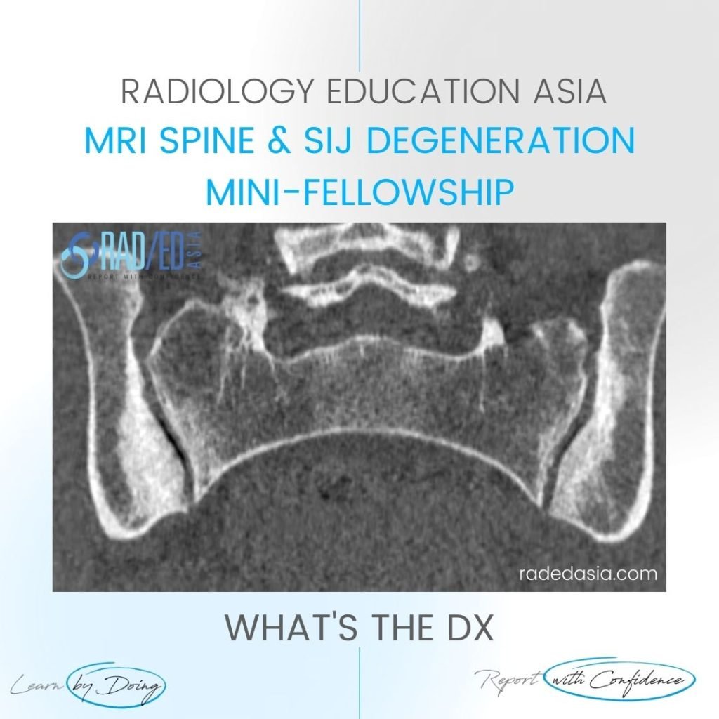
OSTEITIS CONDENSANS ILII RADIOLOGY CT SACROILIAC JOINT SIJ DISCUSSION ON OCI Osteitis Condensans Ilii.   Typical CT sclerotic changes of Osteitis Condensans Ilii. The sclerosis is continuous with the SIJ margin and particularly on the patient’s right it has a triangular appearance.   There is also mild sclerosis in the right sacrum adjacent to the …

OSTEITIS CONDENSANS ILII RADIOLOGY CT SACROILIAC JOINT SIJ (VIDEO) Read More »

tibialis posterior tendinitis tendinopathy tear mri radiology

TIBIALIS POSTERIOR TENDINITIS TENDINOPATHY TEARS RADIOLOGY MRI (VIDEO)

TIBIALIS POSTERIOR TENDINITIS TENDINOPATHY TEARS RADIOLOGY MRI WHAT ARE THE FINDINGS The tibialis posterior tendon is enlarged and increased in signal. DIAGNOSIS & DISCUSSION TIBIALIS POSTERIOR TENDINOPATHY AND PARTIAL TEAR. Tibialis posterior tendinopathy and delamination / partial tears. The normal tibialis posterior tendon should be uniformly low signal. Enlargement and or increased intermediate signal indicates …

TIBIALIS POSTERIOR TENDINITIS TENDINOPATHY TEARS RADIOLOGY MRI (VIDEO) Read More »

This site is intended for Medical Professionals only. Use of this site is governed by our Terms of Service and Privacy Statement which can be found by clicking on the links. Please accept before proceeding to the website.
error: Right Click not available.