HIP PARALABRAL CYST MRI LABRUM TEAR DEGENERATION
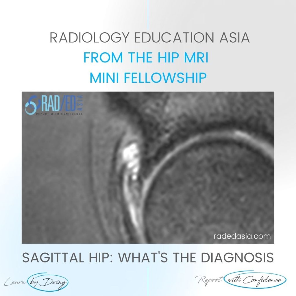
HIP PARALABRAL CYST MRI LABRUM TEAR DEGENERATION DISCUSSION Hip Paralabral cysts are Hyperintense on PD or T2 Fat Sat and can lie adjacent to or track away from the labrum. The presence of a paralabral cyst is always an indicator of an underlying labral tear even if you cant see the tear. The paralabral cyst […]
HIP PARALABRAL CYST MRI LABRUM TEAR DEGENERATION Read More »