
Anterior Cruciate Ligament



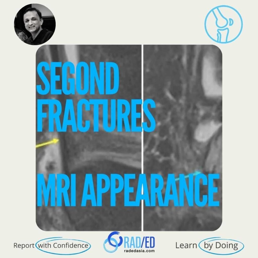

Segond Fractures are fractures of the lateral tibial plateau often associated with ACL tears. It’s thought to be related to the tibial insertion of the anterolateral ligament. However, this ligament is not distinctly identified on MRI, and the best landmark is to look immediately posterior to the ITB insertion.

Lateral tibial plateau, just posterior to the attachment of the ilio-tibial band.
It’s thought to be related to the tibial insertion of the anterolateral ligament. However, this ligament is not distinctly identified on MRI, and the best landmark is to look immediately posterior to the ITB insertion.
Small avulsion/ flake type fracture of the lateral tibial plateau.
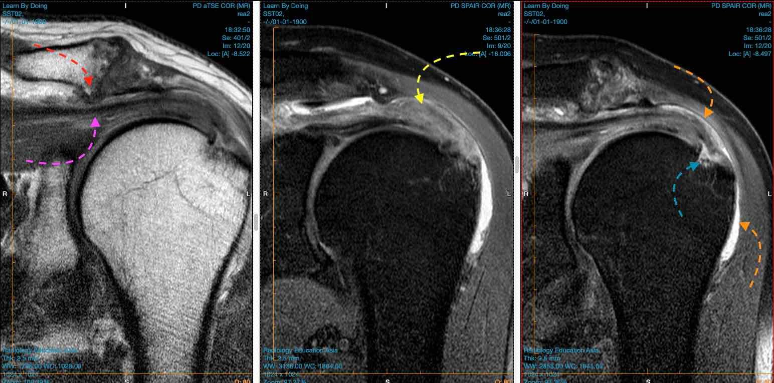

Image Above: X-ray shows fracture fragment (blue arrow)) adjacent to lateral tibial plateau. ITB (yellow arrow) is intact and normal. MRI third image Bone marrow oedema (blue arrow) low signal fracture line seen immediately posterior to ITB insertion.

Image Above: Segond Fracture normal ITB insertion (yellow arrow). Fracture of the lateral cortex (blue arrows) posterior to ITB insertion.
A Segond fracture is an avulsion fracture of the lateral tibial plateau at the site of attachment of the Anterior Longitudinal Ligament (ALL). It is strongly associated with an underlying anterior cruciate ligament (ACL) tear.
Key MRI findings include a a step or break in the black cortical line, bony avulsion fragment at the lateral tibial plateau and in the acute stage bone marrow edema.
A Segond fracture typically results from internal rotation and varus stress to the knee. This mechanism puts excessive tension on the lateral capsular ligament, leading to the avulsion at the site of attachment of the Anterior Longitudinal Ligament (ALL).