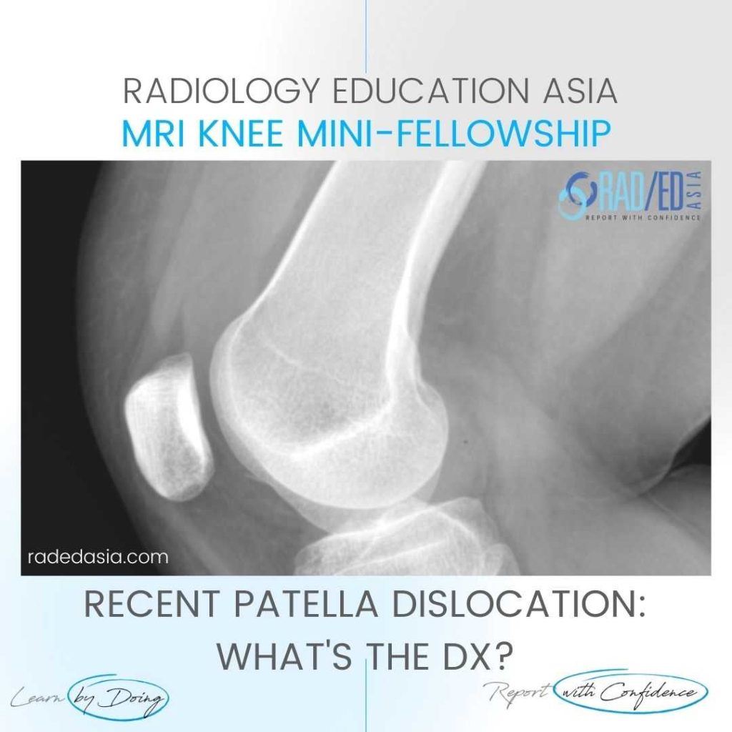
- The patient has had a recent patella dislocation and there is now a large suprapatellar effusion (on MRI this was a hemarthrosis).
- On X-ray we can't say defnitely that it's a hemarthrosis but the presence of a fracture would indicate that it is likely to be a hemarthrosis.
- To diagnose a lipohemarthrosis on X-ray you need to do a horizontal view with the patient lying down in order to see the fat fluid level. Not done in this case.
- On X-ray we can't say defnitely that it's a hemarthrosis but the presence of a fracture would indicate that it is likely to be a hemarthrosis.
- Additionally a fracture fragment is present adjacent to the medial femoral condyle
- This is due to the dislocation, with the patella hitting against the femur to cause the fracture.
- Its not common to see femoral fractures with a patella dislocation and this indicates how forceful the dislocation must have been.
![]()
- Suprapatella effusion (Pink arrow).
- Medial femoral condyle fracture (Green Arrow).
![]()