
It doesn’t make sense because air should be low signal on T2 and T1. However if the patient has been lying supine for a few hours to days before the scan (not uncommon if the patient is in hospital), fluid can accumulate in the pocket of gas and this then becomes high signal on T2 and can mimic infection.
The high T2 signal is usually an isolated finding so there should be no other features of discitis/ osteomyelitis like endplate erosion/ destruction or an epidural collection. If a prior CT is available look for the presence of gas in the disc.
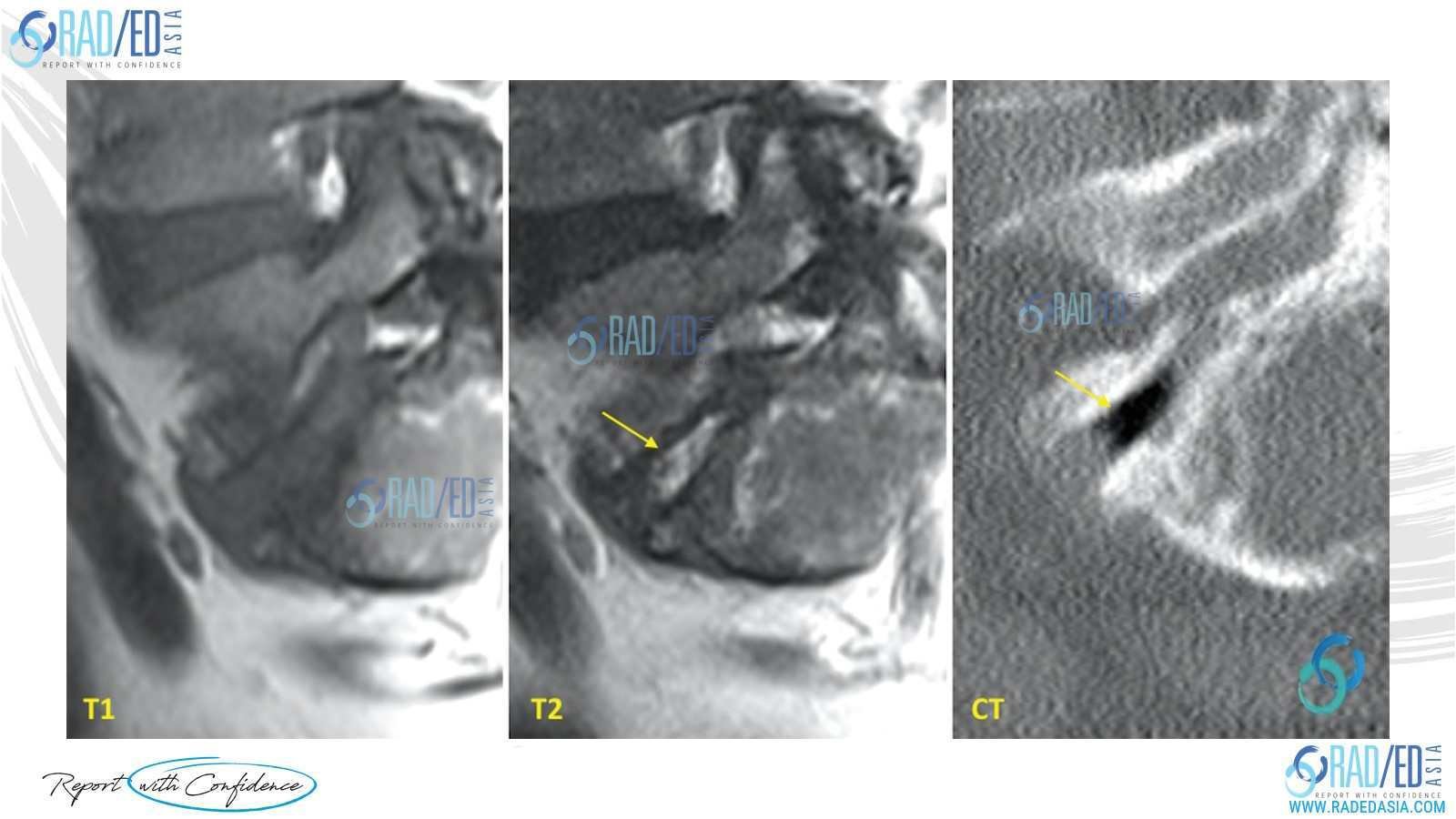
Image 1 above: High T2 signal in the disc mimicking appearance and raising concern of discitis. CT performed same patient a week previously demonstrates gas in the disc in the same region.
Not all areas of vacuum phenomenon will demonstrate increased T2 signal as it depends on whether enough fluid has accumulated in them or not.

Image 2 above: Same Patient as Image 1. Smaller areas of vacuum phenomenon but no increased T2 signal.

Image 3 above: Prior CT demonstrates vacuum phenomenon at L2/3, L3/4 and L4/5 (Blue arrows) with corresponding thin increased T2 signal in degenerate discs (Yellow arrows).
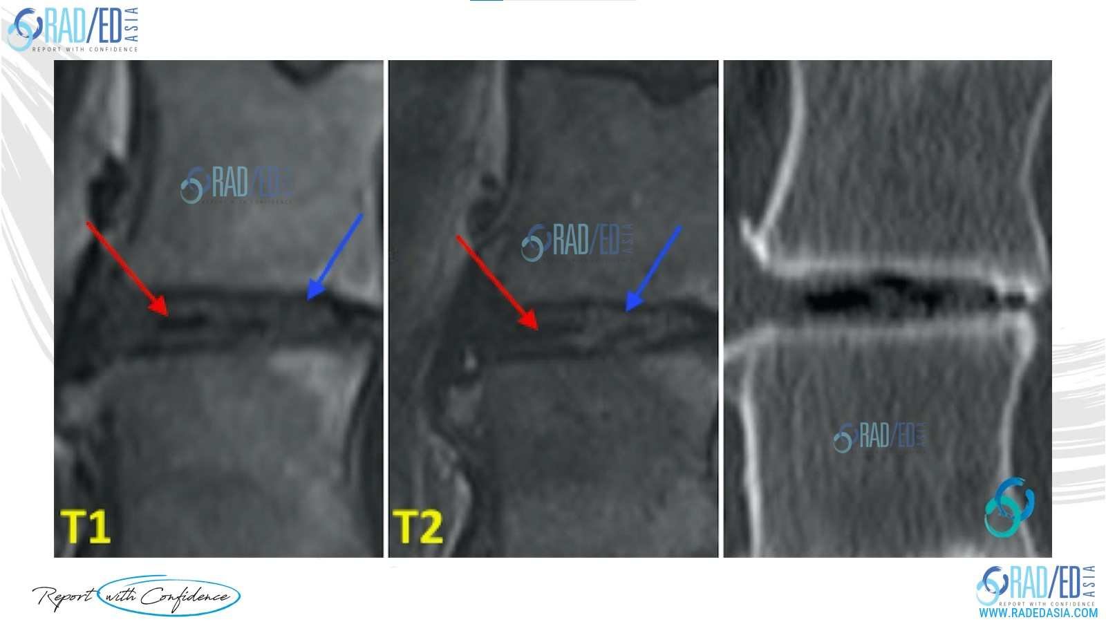
Image 4 above: Magnified view of Image 3 demonstrates a mixture of fluid (Blue arrow) and air (Red arrow) in the disc.
- Sometime Modic Type 1 changes (endplate oedema) may be present together with the T2 disc signal. This can make things more difficult to interpret because the combination of endplate oedema and high T2 disc signal becomes even more concerning for early discitis.
- Again, there should be no endplate destruction/ erosion or collections. If a CT or plain x-ray are available check them to see if gas was present in the disc.
- If there wasn't gas or you don't have a prior CT to review, then it becomes very difficult to differentiate from early discitis and correlation with inflammatory markers and a repeat study to assess for progression is required.

Image 5 above: Same patient as Image 3. Modic Type 1 changes with endplate oedema (Green arrows) in combination with high T2 signal in disc and vacuum phenomenon on CT.


#radedasia #mri #mskmri #radiology