POST OP MRI SPINE ONLINE RADIOLOGY COURSE
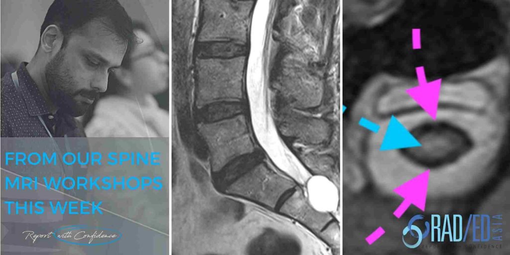
KEY POINTS FROM OUR SPINE MRI COURSES Post Operative Spine MRI Online Radiology Course What are some of the normal post operative changes at the disc on MRI. Another three quick images with some basic key points from our online spine MRI courses. POST OP DISC T2 HYPERINTENSITY Post operative discs can demonstrate hyperintense …