sacral fracture lumbar mri radiology education asia

When the clinical examination and referral notes are inadequate (Back Pain F.I.) the cause of the pain may well be sacral rather than lumbar.

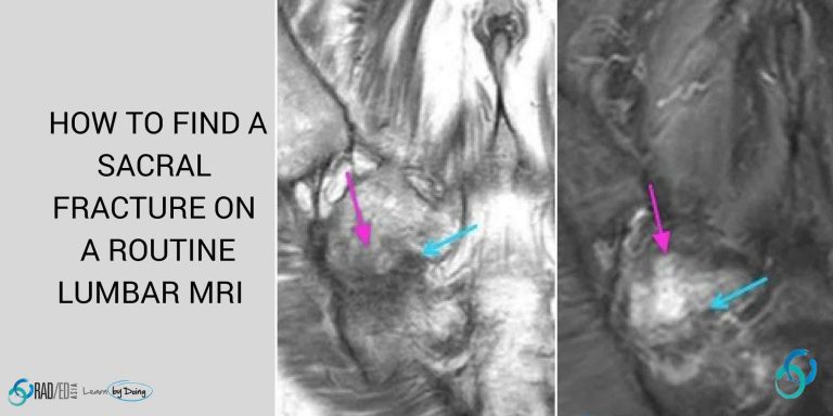
On a standard lumbar spine MRI with axial and sagittal scans,  the sacrum is not specifically imaged, but sacral fractures can be a source of back pain.

There are 2 things you can do to find a sacral fracture which may be the cause of the patient's back pain?

The Best Way is to routinely include a coronal larger FOV T2 Fat Sat or STIR scan of the lumbar spine and sacrum in your sequences. This will demonstrate oedema and the fracture line and makes reporting life a lot easier.


sacrum fracture mri fellowship radiology education asia

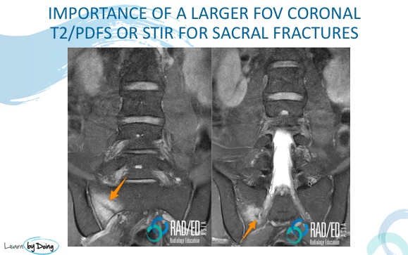
Image Above: Larger FOV T2FS easily demonstrates a sacral fracture which could be missed on standard sagittal and axial lumbar T1 and T2 MRI.

  • The other way if you cant convince people to add an extra sequence.


  • The problem with Lumbar scan sagittals is that they don't go lateral enough to see the entire sacrum (see the coronal image below where the yellow line indicates the lateral most scan).


  • The key then is to look at the lateral most scans that you have on both sides of mid line and look for signal abnormality on the T1 scans.


  • What you are looking for is reduced T1 marrow signal that is an indicator of oedema.


  • T2 scans are not as useful as on the T2 scans the oedema is not as obvious as often its not much brighter than adjacent fat.


  • You will see this only on the lateral most scans as usually the oedema doesn't spread to the mid line scans.


sacral ala fracture mri raded asia radiology education


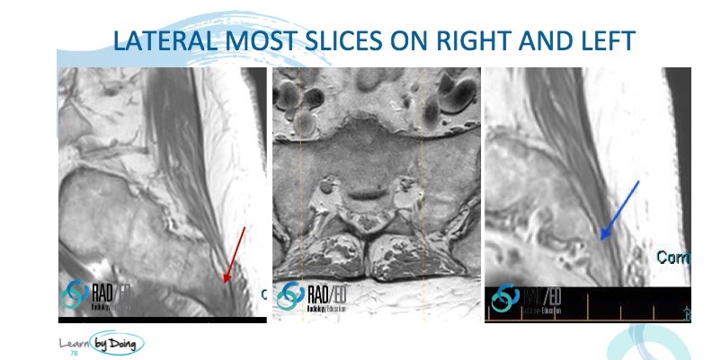
Image Above: Red arrow lateral most scan right of mid line and blue arrow lateral most scan left of mid line. Reduced T1 marrow signal on right compared to left is the clue to a sacral abnormality. Central image with dashed yellow lines demonstrates level of lateral most scans.

The image below is from the same patient demonstrating the sacral fracture on the coronal scan  and the corresponding lateral most scan on the patient's right which demonstrates  low marrow signal on T1.


mri sacral ala fracture raded asia radiology education

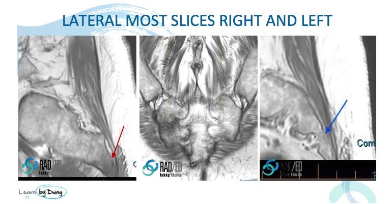
Image above: Red Arrow reduced T1 signal on right side lateral most scan. Compare with normal T1 signal on left (blue arrow). Central image demonstrates the right sacral fracture dashed yellow line indicates location of lateral most sagittal scan on the patient's right.


If you hadn't seen the low T1 marrow signal on the sagittal scan you wouldn't have been able to get a coronal scan to further assess it and diagnose the fracture.

  • Adding a routine larger FOV  coronal STIR or T2FS to the lumbar series makes the diagnosis much easier and is what I would recommend.


  • But when you don't have it, make sure you look at the lateral most slices for reduced T1 marrow signa which is your clue for an abnormality.

#radiology #radiologist #radiologia #mri #spinemri #fracture #lumbarmri #medicalimaging #imaging #futureradiologist #radiologyresident #foamrad #frcr #ortho #ct #emergencydepartment #orthopaedics #radiologylife #trauma #radiologystudent #mskrad #diagnosticimaging #mskradiology #radedasia #radiologyeducation #radiologycases #radiologycme #rheumatology #arthritis #rheumatologist #degenerativedisease #painphysician #chiropractic #physiotherapy #sportsmed

#radedasia #mri #mskmri #radiology

This site is intended for Medical Professionals only. Use of this site is governed by our Terms of Service and Privacy Statement which can be found by clicking on the links. Please accept before proceeding to the website.
error: Right Click not available.