fracture

weight bearing x-rays for lisfranc joint injury instability fracture radedasia

WEIGHT BEARING X-RAYS FOR LISFRANC JOINT INJURY INSTABILITY & FRACTURE (VIDEO)

THE MAIN POINTS TO NOTE OVERVIEW WEIGHT BEARING X-RAYS FOR LISFRANC JOINT INJURY INSTABILITY & FRACTURE   Early diagnosis of Lisfranc instability is crucial to prevent premature OA in the mid foot. Subtle instability patterns can be very difficult to identify on xrays.  Plain radiographs may not effectively identify instability at the LisFranc Joint, especially …

WEIGHT BEARING X-RAYS FOR LISFRANC JOINT INJURY INSTABILITY & FRACTURE (VIDEO) Read More »

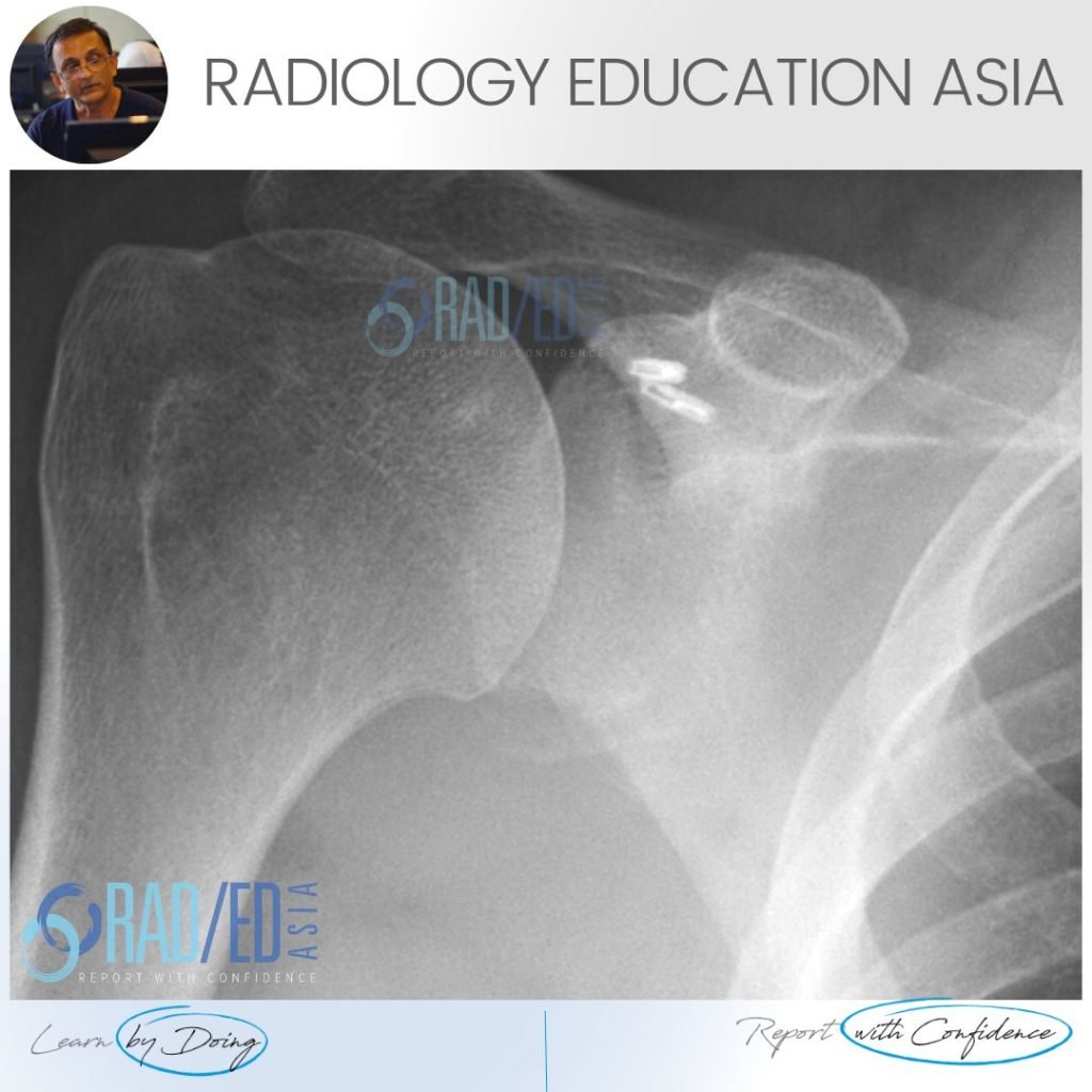
bony bankart xray ct radiology glenoid fracture shoulder dislocation

SHOULDER DISLOCATION BONY BANKART GLENOID FRACTURE XRAY CT (VIDEO)

BONY BANKART LESION RADIOLOGY XRAY FINDING 1: BONE FRAGMENT Image Above: There is a thin fragment of bone at the inferior margin of the glenoid. FINDING 2: GLENOID FLATTENING Image Above: The inferior margin of the glenoid should normally be rounded and ovoid. Here it is flattened which is abnormal. DISCUSSION Bony Bankart lesions on …

SHOULDER DISLOCATION BONY BANKART GLENOID FRACTURE XRAY CT (VIDEO) Read More »

subcortical subchondral fracture knee tibial plateau mri insufficiency

SUBCORTICAL SUBCHONDRAL FRACTURE KNEE MRI TIBIAL PLATEAU (VIDEO)

SUBCORTICAL SUBCHONDRAL FRACTURE KNEE MRI TIBIAL PLATEAU DISCUSSION Subchondral or subcortical fractures of the knee can occur in the femoral condyles or tibial plateau.   The key to the diagnosis is seeing a linear low signal line adjacent to and paralleling the cortex without any cortical defect or break in the acute phase.     There …

SUBCORTICAL SUBCHONDRAL FRACTURE KNEE MRI TIBIAL PLATEAU (VIDEO) Read More »

spine trauma imaging facet fracture dislocation radiology ct

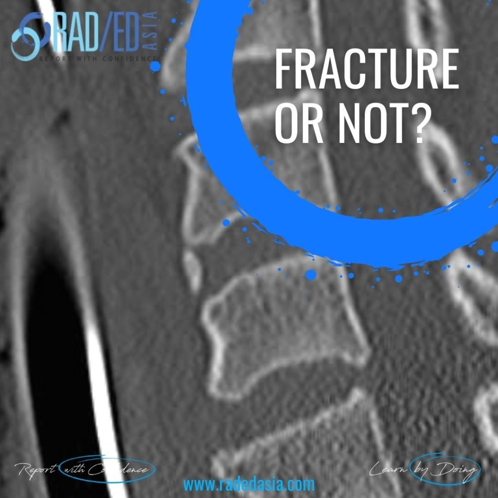
FACET FRACTURE SPINE RADIOLOGY CT TRAUMA IMAGING

FACET FRACTURE RADIOLOGY CT DISCUSSION FACET FRACTURE CT APPEARANCE The facet is fractured (Pink arrows) but there is no evidence on this image of facet subluxation or dislocation.   However the facet joint is widened inferiorly (Green arrow) indicating that the facet capsule must be torn. FACET FRACTURE RADIOLOGY CT: SLIDER TO VIEW IMAGES ARE …

FACET FRACTURE SPINE RADIOLOGY CT TRAUMA IMAGING Read More »

This site is intended for Medical Professionals only. Use of this site is governed by our Terms of Service and Privacy Statement which can be found by clicking on the links. Please accept before proceeding to the website.
error: Right Click not available.