WTD

medullary infarct radiology femur tibia mri radedasia

MEDULLARY BONE INFARCTS RADIOLOGY KNEE MRI

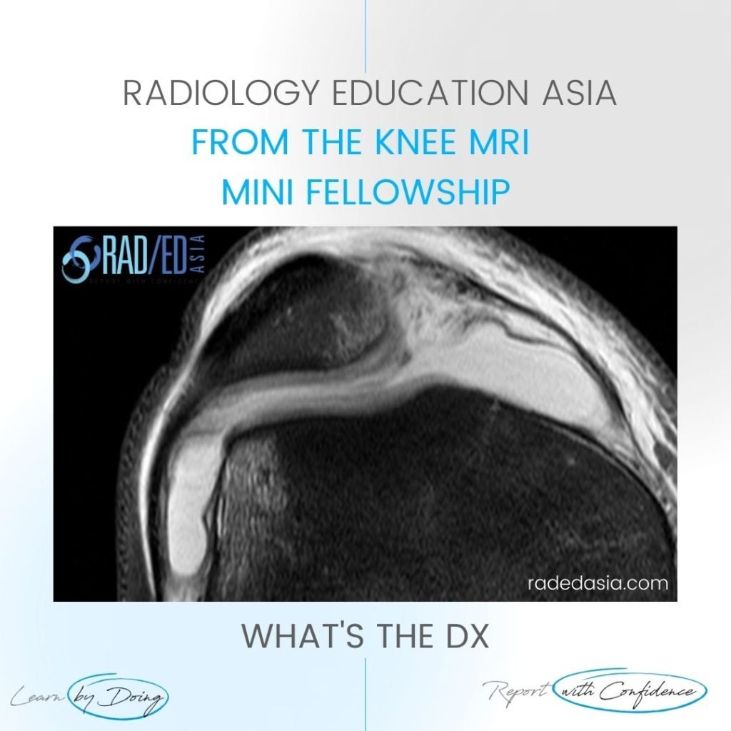
MOVE SLIDER ICON (ARROWS) LEFT TO SEE THE DIAGNOSIS FINDINGS Bilateral medullary femoral and tibial serpiginous STIR hyper-intensity with extension into the femoral and tibial epiphysis. DIAGNOSIS MEDULLARY BONE INFARCTS. This is a characteristic appearance of medullary bone infarcts. Most commonly they are in the diaphysis and metaphysis but in this case they also extend …

MEDULLARY BONE INFARCTS RADIOLOGY KNEE MRI Read More »

learn shoulder mri biseps disloc cyst radedasia

BICEPS DISLOCATION MRI SHOULDER RADIOLOGY

BICEPS DISLOCATION MRI SHOULDER RADIOLOGY DISCUSSION ON BICEPS DISLOCATION Extra articular Biceps dislocation. This is an Extra articular Biceps dislocation into a partial tear the Subscapularis tendon. FINDINGS IN BICEPS DISLOCATION Empty Bicipital Groove (Blue arrow).   Biceps tendon dislocated (Pink arrow).   Partial tear of Subscapularis tendon (Green arrow). High signal is seen within the …

BICEPS DISLOCATION MRI SHOULDER RADIOLOGY Read More »

ankle mri synovitis capsulosynovitis

ANKLE SYNOVITIS MRI RADIOLOGY

ANKLE SYNOVITIS MRI RADIOLOGY DISCUSSION ON ANKLE SYNOVITIS Capsulosynovitis. The normal capsule is thin, well defined and low signal. FINDINGS IN ANKLE SYNOVITIS Thickening and hyper-intensity of the anterior ankle capsule/ synovium. ANKLE SYNOVITIS MRI RADIOLOGY: MOVE SLIDER TO VIEW IMAGES ARE FROM OUR INSTAGRAM ACCOUNT. CLICK ICON BELOW TO SEE ALL OUR INSTAGRAM POSTS …

ANKLE SYNOVITIS MRI RADIOLOGY Read More »

mri hip cam fai femoro acetabular impingement rea

MRI HIP CAM FAI FEMORO ACETABULAR IMPINGEMENT

MRI HIP CAM FAI FEMORO ACETABULAR IMPINGEMENT MRI CAM LESION FAI DISCUSSION This is a CAM type lesion / deformity of the femoral neck. There is convexity of the supero lateral femoral head neck junction. The normal supero lateral femoral head neck junction should be concave like the inferior junction. The convexity may be diffuse …

MRI HIP CAM FAI FEMORO ACETABULAR IMPINGEMENT Read More »

mri shoulder ligaments mghl middle glenohumeral ligament radedasia

SHOULDER MRI MIDDLE GLENO HUMERAL LIGAMENT

MIDDLE GLENO HUMERAL LIGAMENT MRI SHOULDER MGHL DISCUSSION ON MGHL This is the normal MGHL (Middle Gleno Humeral Ligament). It lies adjacent and anterior to the labrum (Green arrow) and extends inferiorly to join the anterior capsule (Blue arrow) deep to subscapularis. FINDINGS IN MGHL Well defined low signal structure (Pink arrows) adjacent to the …

SHOULDER MRI MIDDLE GLENO HUMERAL LIGAMENT Read More »

mri knee course patella dislocation

KNEE MRI PATELLA DISLOCATION SUBLUXATION

KNEE MRI PATELLA DISLOCATION SUBLUXATION DISCUSSION PATELLA DISLOCATION Patella Dislocation Subluxation MRI.  There has been a recent Patella dislocation with classical contra coup bone bruising and torn MPFL/ medial retinaculum.   The best clue to patella dislocation / subluxation on MRI is the presence of the contra coup bone bruising in the patella and femur. …

KNEE MRI PATELLA DISLOCATION SUBLUXATION Read More »

mri ankle os peroneum learn radiology wtd radedasia

ANKLE MRI OS PERONEUM: WHAT IS THE DX

MOVE SLIDER ICON (ARROWS) LEFT TO SEE THE DIAGNOSIS FINDINGS Well corticated ossified structure adjacent to the calcaneo cuboid articulation.. DIAGNOSIS Os peroneum.Ossicle within the Peroneus Longus Tendon at the level of the Calcaneo Cuboid joint. Can be single, bipartite or multipartite. IMAGES ARE FROM OUR INSTAGRAM ACCOUNT. CLICK ICON BELOW TO SEE ALL OUR …

ANKLE MRI OS PERONEUM: WHAT IS THE DX Read More »

This site is intended for Medical Professionals only. Use of this site is governed by our Terms of Service and Privacy Statement which can be found by clicking on the links. Please accept before proceeding to the website.
error: Right Click not available.