WTD

freiberg disease mri radiology toe radedasia

FREIBERG DISEASE SUBCHONDRAL FRACTURE INFRACTION OSTEOCHONDROSIS (VIDEO)

FREIBERG DISEASE / SUBCHONDRAL FRACTURE INFRACTION / OSTEOCHONDROSIS DISCUSSION FREIBERG DISEASE / SUBCHONDRAL FRACTURE INFRACTION / OSTEOCHONDROSIS Extensive oedema in the head of the 2nd metatarsal with slight flattening of the cortical margin is characteristic of what’s called Freiberg’s Disease in adolescents and Subchondral Fracture in adults. Freiberg disease occurs in adolescents with an unfused […]

FREIBERG DISEASE SUBCHONDRAL FRACTURE INFRACTION OSTEOCHONDROSIS (VIDEO) Read More »

spinal epidural lipomatosis mri lumbar thoracic radedasia

SPINAL EPIDURAL LIPOMATOSIS LUMBAR & THORACIC MRI

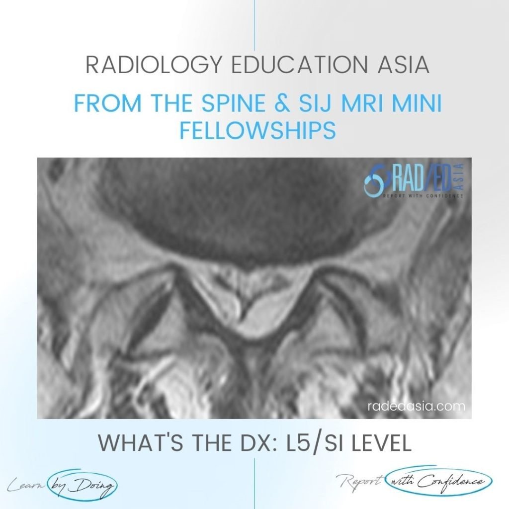
Stay tuned on new Fellowships and learnings Subscribe The Dx / Spine, Spine Degeneration SPINAL EPIDURAL LIPOMATOSIS Spinal epidural lipomatosis is an excessive accumulation of non encapsulated fat in the epidural space. It can be seen on MRI in the thoracic or lumbar spine. In the cervical spine epidural lipomatosis is not seen. Epidural lipomatosis

SPINAL EPIDURAL LIPOMATOSIS LUMBAR & THORACIC MRI Read More »

itb iliotibial band friction syndrome bursa radiology mri knee

ITB ILIOTIBIAL BAND FRICTION SYNDROME MRI: IS IT A BURSA OR A RECESS?

Stay tuned on new Fellowships and learnings Subscribe The Dx / Knee ITB ILIOTIBIAL BAND FRICTION SYNDROME MRI TWO POSSIBILLITIES With fluid deep to the ITB we have 2 possibilities. It’s a recess of the suprapatellar bursa or, It’s an ITB bursa (an adventitial bursa which develops due to friction). HOW DOES THE ANATOMY HELP

ITB ILIOTIBIAL BAND FRICTION SYNDROME MRI: IS IT A BURSA OR A RECESS? Read More »

ankylosing spondylitis spine radiology ct mri fusion images

ANKYLOSING SPONDYLITIS SPINE RADIOLOGY CT MRI FUSION IMAGES

Stay tuned on new Fellowships and learnings Subscribe The Dx / Spine ANKYLOSING SPONDYLITIS SPINE RADIOLOGY CT MRI FUSION WHERE TO LOOK There are 4 sites in the posterior spine to assess for changes of AS. Facet Joints.   Superspinous Ligament.   Costovertebral joints.   Costo transverse joints. Changes can be at 1 or more

ANKYLOSING SPONDYLITIS SPINE RADIOLOGY CT MRI FUSION IMAGES Read More »

radiology knee segond arcuate lateral fibular fracture xray mri knee radedasia

3 LATERAL KNEE FRACTURES SEGOND ARCUATE AND LATERAL FIBULAR FRACTURES XRAY

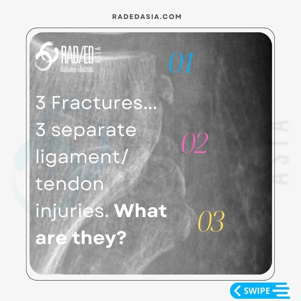
Stay tuned on new Fellowships and learnings Subscribe The Dx / Knee 3 LATERAL KNEE FRACTURE: WHAT THEY ARE AND WHAT THEY MEAN 01: SEGOND FRACTURE • Avulsion Fracture of the lateral tibial plateau.   • Occurs at attachment site of the Antero Lateral Ligament.   • Posterior to IT B insertion site.   •

3 LATERAL KNEE FRACTURES SEGOND ARCUATE AND LATERAL FIBULAR FRACTURES XRAY Read More »

EPIDURAL SPACE INVADERS: SPINE MRI EPIDURAL LIPOMATOSIS PART 2

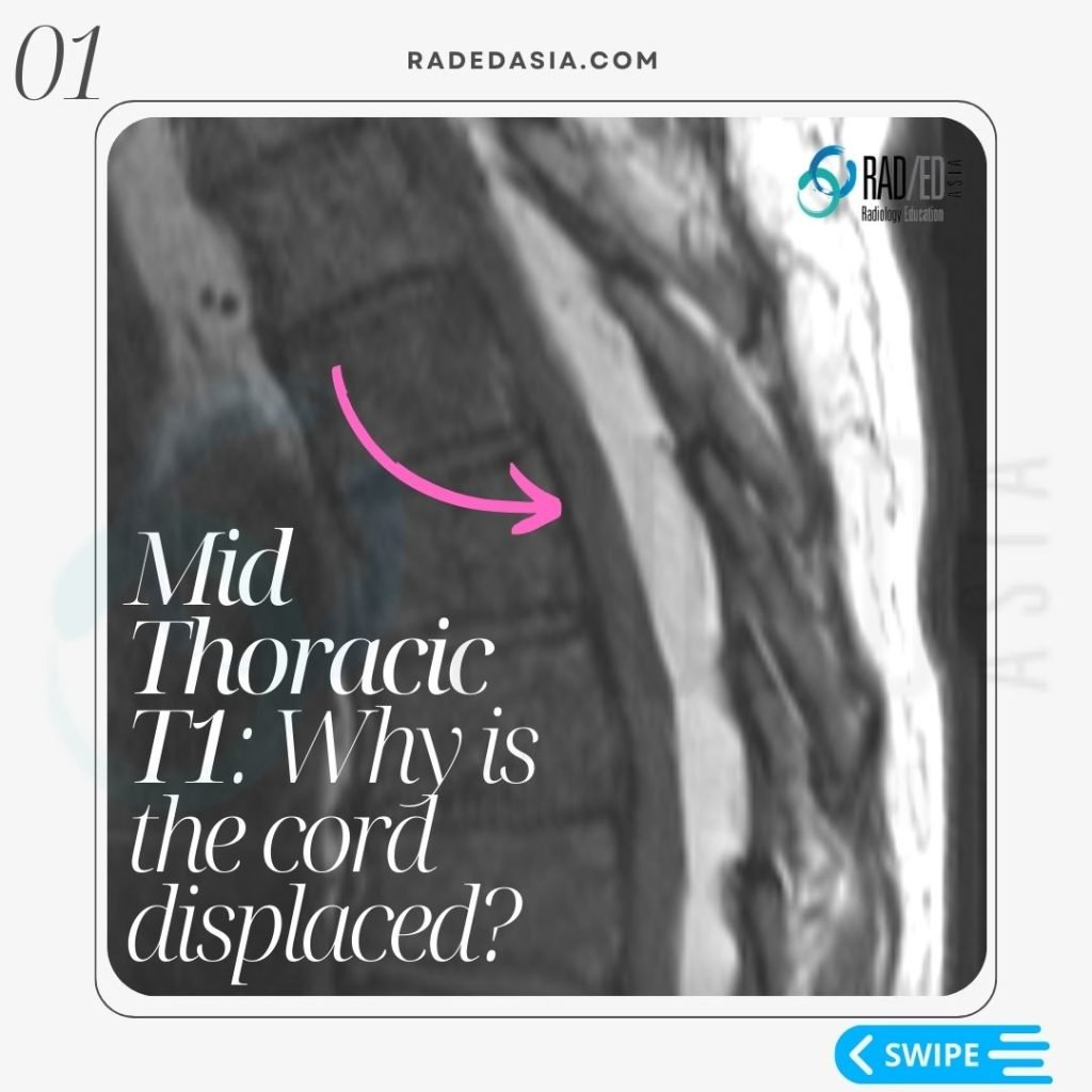
Stay tuned on new Fellowships and learnings Subscribe The Dx / Spine THORACIC EPIDURAL LIPOMATOSIS RADIOLOGY OVERVIEW Thoracic Epidural lipomatosis is not uncommon to see but has a different appearance to Lumbar Epidural Lipomatosis WHAT IS IT? With thoracic epidural lipomatosis there is excess fat accumulated in the epidural space. WHAT ARE THE CAUSES? There

EPIDURAL SPACE INVADERS: SPINE MRI EPIDURAL LIPOMATOSIS PART 2 Read More »

meniscal tear radiology mri knee radedasia

MENISCUS TEAR MRI KNEE: MISSING MENISCI WHAT TO LOOK FOR

Stay tuned on new Fellowships and learnings Subscribe The Dx / Knee MENISCUS TEAR MRI KNEE WHAT TO LOOK FOR In a Normal meniscus the superior and inferior halves should be symmetric. In all three cases there is localised loss of ” volume” of the meniscus. This gives the meniscus an asymmetric look. Asymmetry in

MENISCUS TEAR MRI KNEE: MISSING MENISCI WHAT TO LOOK FOR Read More »

knee mri bursa suprapatellar plica

KNEE MRI PLICA SUPRAPATELLAR BURSA LIPOHEMARTHROSIS

Stay tuned on new Fellowships and learnings Subscribe The Dx / Knee KNEE MRI PLICA SUPRAPATELLAR BURSA LIPOHEMARTHROSIS NORMAL SUPRA PATELLAR PLICA The normal supra patellar plica is not continuous.   There is a gap in it ( fenestration) which allows fluid to circulate freely in the SP bursa above and below the plica. CONTINUOUS

KNEE MRI PLICA SUPRAPATELLAR BURSA LIPOHEMARTHROSIS Read More »

knee-infrapatellar-plica-syndrome-radiology-mri-radedasia-1

KNEE INFRAPATELLAR PLICA SYNDROME RADIOLOGY MRI (VIDEO)

KNEE INFRAPATELLAR PLICA SYNDROME RADIOLOGY MRI DISCUSSION ON KNEE INFRAPATELLAR PLICA PLICAS IN THE KNEE The infrapatellar plica is one of 4 plicas in the knee: Medial Patella Plica. Suprapatellar Plica. Infrapatellar plica. Lateral Patellar plica. INFRAPATELLAR PLICA ANATOMY Attaches to anterior intercondylar notch and inferior pole patella. Runs through Hoffa’s Fat Pad. In the

KNEE INFRAPATELLAR PLICA SYNDROME RADIOLOGY MRI (VIDEO) Read More »

error: Content is protected !!

Join our Newsletter

Stay tuned on new
Mini-Fellowships launches and learnings

Mini-Fellowships

Better Than Conferences

Join our WhatsApp Community

We help radiologists learn
by doing and report with confidence

This site is intended for Medical Professions only. Use of this site is governed by our Terms of Service and Privacy Statement which can be found by clicking on the links. Please accept before proceeding to the website.