KNEE

LEARN MSK MRI KNEE ONLINE     knee icon radedasia

KNEE MSK MRI RADIOLOGY: We regularly post on Knee MRI and our Knee MRI Courses with all the posts on this page.

Knee MRI can be difficult due to the complex anatomy. In our posts we have concise explanation of the Knee MRI anatomy, the macroscopic pathology that’s relevant to the Imaging and the Imaging findings in various pathologies.

Learn where to Look, What to Look for and How to Report Knee MRI More Confidently

meniscal tear radiology mri knee radedasia

MENISCUS TEAR MRI KNEE: MISSING MENISCI WHAT TO LOOK FOR

MENISCUS TEAR MRI KNEE WHAT TO LOOK FOR • In a Normal meniscus the superior and inferior halves should be symmetric. • In all three cases there is localised loss of ” volume” of the meniscus. • This gives the meniscus an asymmetric look. • Asymmetry in a meniscus is always abnormal and we need …

MENISCUS TEAR MRI KNEE: MISSING MENISCI WHAT TO LOOK FOR Read More »

knee mri bursa suprapatellar plica

KNEE MRI PLICA SUPRAPATELLAR BURSA LIPOHEMARTHROSIS

KNEE MRI PLICA SUPRAPATELLAR BURSA LIPOHEMARTHROSIS NORMAL SUPRA PATELLAR PLICA The normal supra patellar plica is not continuous.  There is a gap in it ( fenestration) which allows fluid to circulate freely in the SP bursa above and below the plica. CONTINUOUS SUPRAPATELLAR PLICA At times the SP Plica is continuous with no gap.  Normally …

KNEE MRI PLICA SUPRAPATELLAR BURSA LIPOHEMARTHROSIS Read More »

knee infrapatellar plica syndrome radiology mri radedasia

KNEE INFRAPATELLAR PLICA SYNDROME RADIOLOGY MRI (VIDEO)

KNEE INFRAPATELLAR PLICA SYNDROME RADIOLOGY MRI DISCUSSION ON KNEE INFRAPATELLAR PLICA PLICAS IN THE KNEE The infrapatellar plica is one of 4 plicas in the knee: Medial Patella Plica. Suprapatellar Plica. Infrapatellar plica. Lateral Patellar plica. INFRAPATELLAR PLICA ANATOMY Attaches to anterior intercondylar notch and inferior pole patella.   Runs through Hoffa’s Fat Pad.   …

KNEE INFRAPATELLAR PLICA SYNDROME RADIOLOGY MRI (VIDEO) Read More »

osgood schlatter xray radiology radedasia

OSGOOD SCHLATTER RADIOLOGY X-RAY (VIDEO)

OSGOOD SCHLATTER RADIOLOGY X-RAY FINDINGS There are three findings in the x-ray that make the diagnosis ofOsgood Schlatter. Bone avulsion from tibial tuberosity.  Soft tissue swelling over tibial tuberosity.  Increased density deep infra patella bursa from fluid/inflammation. OSGOOD SCHLATTER X-RAY FINDINGS: VIEW VIDEO https://youtu.be/qG4b7ik9G3I If your Browser is blocking the video, Please view it on …

OSGOOD SCHLATTER RADIOLOGY X-RAY (VIDEO) Read More »

msk radiology workshop conference course musculoskeletal mri knee shoulder imaging abu dhabi uae radedasia

MSK RADIOLOGY CONFERENCE COURSE MUSCULOSKELETAL MRI KNEE & SHOULDER ABU DHABI UAE

Home MSK RADIOLOGY CONFERENCE COURSE MUSCULOSKELETAL MRI WORKSHOP UAE ABU DHABI: MSK MRI RADIOLOGY KNEE & SHOULDER MINI FELLOWSHIP: 25/26 FEBRUARY 2023 This Mini Fellowship is now over.To see our latest Onsite Guided Mini Fellowships please click HERE WHAT HAPPENED AT THE MINI-FELLOWSHIP @ ABU DHABI CLICK ON THE IMAGE BELOW TO FIND OUT Our …

MSK RADIOLOGY CONFERENCE COURSE MUSCULOSKELETAL MRI KNEE & SHOULDER ABU DHABI UAE Read More »

subcortical subchondral fracture knee tibial plateau mri insufficiency

SUBCORTICAL SUBCHONDRAL FRACTURE KNEE MRI TIBIAL PLATEAU (VIDEO)

SUBCORTICAL SUBCHONDRAL FRACTURE KNEE MRI TIBIAL PLATEAU DISCUSSION Subchondral or subcortical fractures of the knee can occur in the femoral condyles or tibial plateau.   The key to the diagnosis is seeing a linear low signal line adjacent to and paralleling the cortex without any cortical defect or break in the acute phase.     There …

SUBCORTICAL SUBCHONDRAL FRACTURE KNEE MRI TIBIAL PLATEAU (VIDEO) Read More »

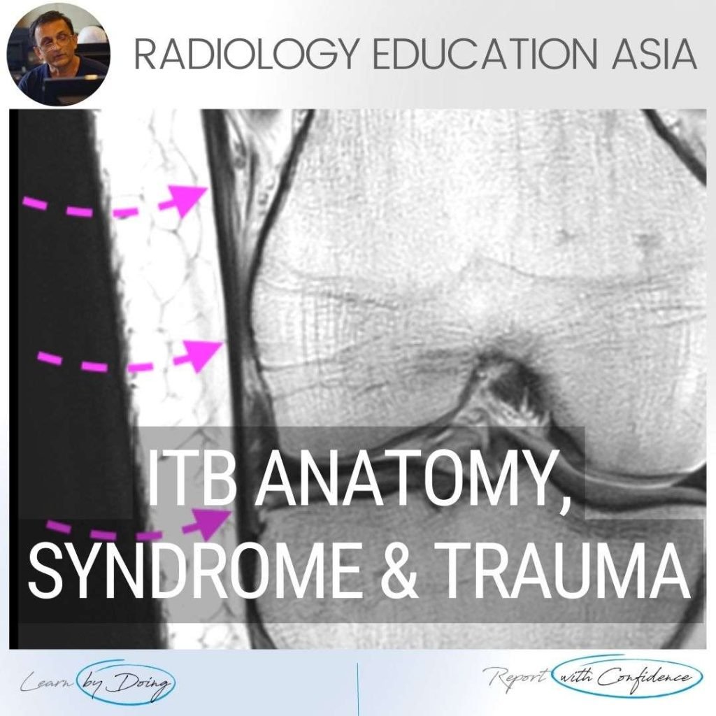
iliotibial band mri anatomy syndrome trauma mri knee radedasia

ILIOTIBIAL BAND MRI ANATOMY, SYNDROME, TRAUMA KNEE RADIOLOGY

ILIOTIBIAL BAND MRI ANATOMY, SYNDROME, TRAUMA Finding and assessing the Iliotibial Band (ITB) on MRI of the Knee is the first structure we assess when looking at the lateral side of the Knee.  In these three posts we look at: The normal MRI anatomy of the ITB. MRI of Iliotibial Band Syndrome (ITB Friction Syndrome) …

ILIOTIBIAL BAND MRI ANATOMY, SYNDROME, TRAUMA KNEE RADIOLOGY Read More »

meniscus tear maceration degeneration mri knee radedasia

MENISCUS TEAR MACERATION DEGENERATION MRI KNEE RADIOLOGY (VIDEO)

MENISCUS MACERATION DEGENERATION MRI KNEE WHAT ARE THE FINDINGS MENISCUS MACERATION DEGENERATION MRI KNEE   Generalized increase in meniscal signal. Intermediate increase in signal on PD and PD/T2FS. The meniscus generally retains it’s shape but is ill defined. Due to the degeneration its more likely to tear and it also stops functioning like a normal …

MENISCUS TEAR MACERATION DEGENERATION MRI KNEE RADIOLOGY (VIDEO) Read More »

This site is intended for Medical Professionals only. Use of this site is governed by our Terms of Service and Privacy Statement which can be found by clicking on the links. Please accept before proceeding to the website.
error: Right Click not available.