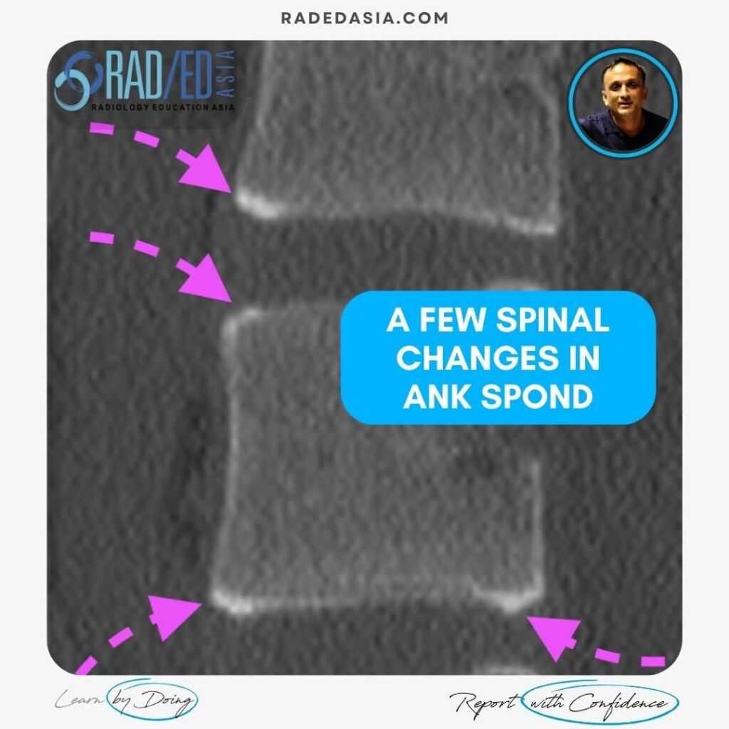
A FEW SPINAL CHANGES IN ANKYLOSING SPONDYLITIS
ANKYLOSING SPONDYLITIS SPINAL CHANGES CT & MRI Findings in Ankylosing Spondylitis of the spine Ankylosing spondylitis (AS), in the spine can involve the anterior and posterior elements. It usually commences anteriorly and as the disease progresses, posterior changes are seen. In these images we look at: Corner erosions and Sclerosis. Syndesmophyte formation between vertebrae. Fusion: …