MRI

CEREBELLAR INFARCTS: HOW NOT TO MISS THEM

The Problem: Acute cerebellar infarcts are easy to see on diffusion imaging, but small chronic infarcts in the cerebellum are often missed because they are confused with normal cerebellar sulci. How do you differentiate small chronic cerebellar infracts from normal sulci? The Answer: In the Cerebellar Hemispheres: Normal: Chronic Infarct: Infarcts usually run perpendicular to the …

CEREBELLAR INFARCTS: HOW NOT TO MISS THEM Read More »

cavernoma-cord-mri-radiology-education

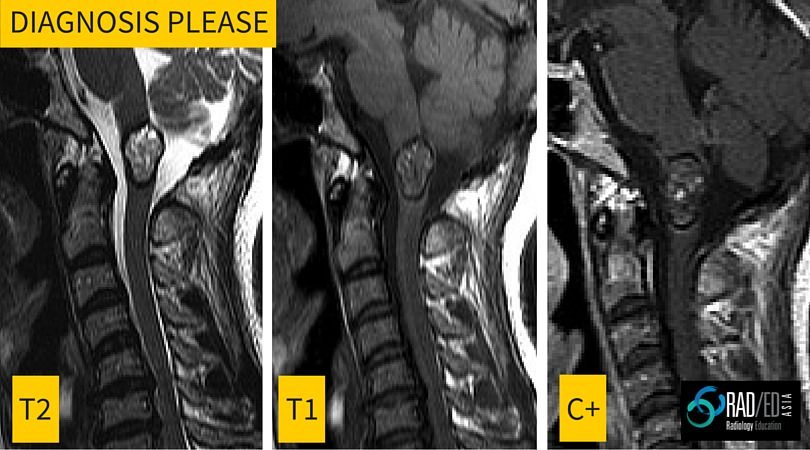
Radiology Education Diagnosis Please: Spinal cord lesion MRI

This is the first in a regular series of posts on cases of interest. Incidental finding in a 40yo male. Image above.     ANSWER Cavernomas of the spinal cord have a very specific appearance on MRI with the following features Lobulated well defined lesion mixed signal T1 and T2 No significant enhancement Low signal …

Radiology Education Diagnosis Please: Spinal cord lesion MRI Read More »

glenoid-mri-normal-variant-radedasia

GLENOID LABRUM MRI SIMPLIFIED 2: Are you normal or not?

The problem: Many normal labral variants get called tears. How do we differentiate a normal labral variant from a tear? The solution: Normal labral variants occur in specific areas of the glenoid labrum. Understand their typical location and you will not confuse them with tears. If you havent read the first post on the classification …

GLENOID LABRUM MRI SIMPLIFIED 2: Are you normal or not? Read More »

glenoid-mri-labrum-radedasia

GLENOID LABRUM MRI SIMPLIFIED PART 1: There’s a Pear in There

The problem: A lot of the difficulty people have in reporting shoulder MRI and particularly the glenoid labrum is in having a clear understanding of the MRI anatomy. This leads to confusion in differentiating normal glenoid labrum variants from labral tears and SLAP tears. The solution: The humble pear. Only a few dollars a kilo …

GLENOID LABRUM MRI SIMPLIFIED PART 1: There’s a Pear in There Read More »

SWI OR T2*: WHICH MRI SEQUENCE TO USE IN THE DETECTION OF CEREBRAL MICROBLEEDS?

Quick Journal Review: Should you use T2* or SWI sequences to detect cerebral microbleeds.Which sequence is more sensitive? Journal Article: SWI or T2*: Which MRI Sequence to Use in the Detection of Cerebral Microbleeds? The Karolinska Imaging Dementia Study S. Shams et al AJNR 36:1089–95 Jun 2015 Intro: Different sequences can be used to detect …

SWI OR T2*: WHICH MRI SEQUENCE TO USE IN THE DETECTION OF CEREBRAL MICROBLEEDS? Read More »

This site is intended for Medical Professionals only. Use of this site is governed by our Terms of Service and Privacy Statement which can be found by clicking on the links. Please accept before proceeding to the website.
error: Right Click not available.