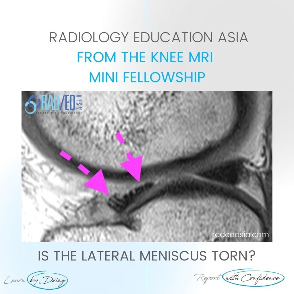
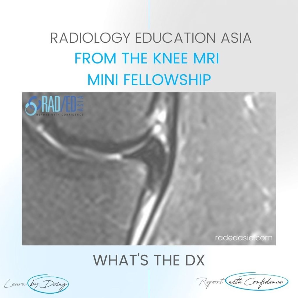
MENISCAL OSSICLES: POSTERIOR HORN MENISCUS PART 3
MENISCAL OSSICLES: POSTERIOR HORN MENISCUS MENISCAL OSSICLES: WHAT ARE THEY Meniscal ossicles are areas of ossification that occur within the meniscus. WHY DO THEY OCCCUR Two possible causes are given: They are a Result of Post traumatic metaplasia and heterotopic ossification of a meniscal tear (either posterior horn or root). This is the most likely …
MENISCAL OSSICLES: POSTERIOR HORN MENISCUS PART 3 Read More »