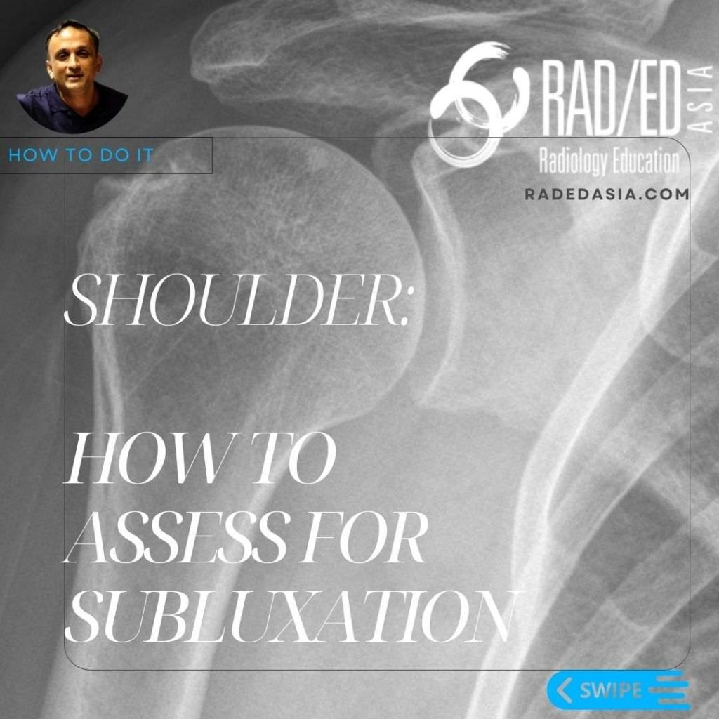
Dr Ravi Padmanabhan

ankylosing spondylitis spine radiology ct mri fusion images

ANKYLOSING SPONDYLITIS SPINE RADIOLOGY CT MRI FUSION IMAGES

ANKYLOSING SPONDYLITIS SPINE RADIOLOGY CT MRI FUSION WHERE TO LOOK There are 4 sites in the posterior spine to assess for changes of AS. Facet Joints.  Superspinous Ligament.  Costovertebral joints.  Costo transverse joints. Changes can be at 1 or more of these sites. THE TAKE AWAY Spinal changes start anteriorly and progress posteriorly.   All …

ANKYLOSING SPONDYLITIS SPINE RADIOLOGY CT MRI FUSION IMAGES Read More »

radiology knee segond arcuate lateral fibular fracture xray mri knee radedasia

3 LATERAL KNEE FRACTURES SEGOND ARCUATE AND LATERAL FIBULAR FRACTURES XRAY

3 LATERAL KNEE FRACTURE: WHAT THEY ARE AND WHAT THEY MEAN 01: SEGOND FRACTURE • Avulsion Fracture of the lateral tibial plateau.  • Occurs at attachment site of the Antero Lateral Ligament.  • Posterior to IT B insertion site.  • Associated with ACL tears. 02: ARCUATE FRACTURE • Fracture of the Fibula Styloid (tip of …

3 LATERAL KNEE FRACTURES SEGOND ARCUATE AND LATERAL FIBULAR FRACTURES XRAY Read More »

spine-thoracic-epidural-lipomatosis- radiology-spinal-spine-mri-radedasia

EPIDURAL SPACE INVADERS: SPINE MRI EPIDURAL LIPOMATOSIS PART 2

THORACIC EPIDURAL LIPOMATOSIS RADIOLOGY OVERVIEW Thoracic Epidural lipomatosis is not uncommon to see but has a different appearance to Lumbar Epidural Lipomatosis WHAT IS IT? With thoracic epidural lipomatosis there is excess fat accumulated in the epidural space. WHAT ARE THE CAUSES? There is an increased risk of developing epidural lipomatosis with: Chronic corticosteroid use. …

EPIDURAL SPACE INVADERS: SPINE MRI EPIDURAL LIPOMATOSIS PART 2 Read More »

meniscal tear radiology mri knee radedasia

MENISCUS TEAR MRI KNEE: MISSING MENISCI WHAT TO LOOK FOR

MENISCUS TEAR MRI KNEE WHAT TO LOOK FOR • In a Normal meniscus the superior and inferior halves should be symmetric. • In all three cases there is localised loss of ” volume” of the meniscus. • This gives the meniscus an asymmetric look. • Asymmetry in a meniscus is always abnormal and we need …

MENISCUS TEAR MRI KNEE: MISSING MENISCI WHAT TO LOOK FOR Read More »

knee mri bursa suprapatellar plica

KNEE MRI PLICA SUPRAPATELLAR BURSA LIPOHEMARTHROSIS

KNEE MRI PLICA SUPRAPATELLAR BURSA LIPOHEMARTHROSIS NORMAL SUPRA PATELLAR PLICA The normal supra patellar plica is not continuous.  There is a gap in it ( fenestration) which allows fluid to circulate freely in the SP bursa above and below the plica. CONTINUOUS SUPRAPATELLAR PLICA At times the SP Plica is continuous with no gap.  Normally …

KNEE MRI PLICA SUPRAPATELLAR BURSA LIPOHEMARTHROSIS Read More »

Coracoid Process tendon attachment coracobrachialis short head biceps

CORACOID PROCESS TENDON ATTACHMENT MRI CORACOBRACHIALIS & SHORT HEAD OF BICEPS (VIDEO)

CORACOID PROCESS TENDON ATTACHMENT MRI CORACOBRACHIALIS & SHORT HEAD OF BICEPS HOW TO FIND CORACOBRACHIALIS & SHORT HEAD OF BICEPS:   There are 3 tendons that attach to the coracoid process. Pectoralis Minor. Coracobrachialis and, The short Head of Biceps.   In this video, we look at how to identify on MRI the Coracobrachialis and …

CORACOID PROCESS TENDON ATTACHMENT MRI CORACOBRACHIALIS & SHORT HEAD OF BICEPS (VIDEO) Read More »

This site is intended for Medical Professionals only. Use of this site is governed by our Terms of Service and Privacy Statement which can be found by clicking on the links. Please accept before proceeding to the website.
error: Right Click not available.