SPINE

SPINE MRI Learn Onlinelearn spine mri workshop online

All our posts on Spine MRI Radiology . We regularly post on Spine MRI with all the posts on this page.

Spine MRI Radiology can be difficult due to the complex anatomy and the many pathologies that occur. In our posts we have concise explanation of the Spine MRI anatomy, the macroscopic pathology that’s relevant to the Imaging and the Imaging findings in various pathologies.

Learn where to Look, What to Look for and How to Report More Confidently

rheumatology course meeting iracon india

INDIAN RHEUMATOLOGY ASSOCIATION COURSE MEETING INDIA IRACON ONLINE

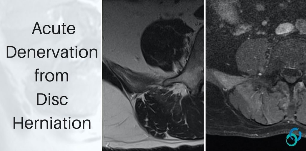
INDIAN RHEUMATOLOGY ASSOCIATION COURSE MEETING INDIA IRACON: MRI SPINE SPONDYLOARTHROPATHY FOR RHEUMATOLOGISTS Dr Ravi spoke at the Indian Rheumatology Association Annual Meeting ( IRACON ) on MRI assessment of the spine in Spondyloarthropathies. This is a talk that is tailored for Rheumatologists.  As a Rheumatologist you are not expected to look at a scan like …

INDIAN RHEUMATOLOGY ASSOCIATION COURSE MEETING INDIA IRACON ONLINE Read More »

fatty filum mri learn spine radiology

MRI FILUM TERMINALE SPINE: WHAT IS THE DX

MOVE SLIDER ICON (ARROWS) LEFT TO SEE THE DIAGNOSIS FINDINGS Bright T1 Linear fatty structure (Pink arrows) in the centre of the canal in the expected location of the filum terminale. DIAGNOSIS FATTY FILUM. Typical appearance of linear T1 hyper-intensity along course of filum terminale. No evidence of tethering of the cord. IMAGES ARE FROM OUR …

MRI FILUM TERMINALE SPINE: WHAT IS THE DX Read More »

denervation lumbar mri paraspinal

MRI LUMBAR: DISC HERNIATION & ACUTE DENERVATION

DISC HERNIATION & ACUTE DENERVATION MRI Lumbar Spine Disc herniation with Paraspinal Muscle Denervation. DISC HERNIATION: WHAT DOES DENERVATION LOOK LIKE Lumbar MRIs are likely the most common MRI people would report but to see acute denervation changes from disc herniation is uncommon. So what does denervation look like in the lumbar spine? ACUTE TO …

MRI LUMBAR: DISC HERNIATION & ACUTE DENERVATION Read More »

romanus lesion endplate anatomy mri enthesitis radedasia header

ROMANUS LESIONS ANATOMY OF THE ENDPLATE AND ENTHESIS

ROMANUS LESIONS ANATOMY OF THE ENDPLATE AND ENTHESIS To understand the Romanus Lesion or Corner Inflammatory Lesion as its described on MRI, its important to understand  the anatomy of the endplate and enthesis it forms with the annulus. This post was done together with Dr Joe Thomas who is a very talented Rheumatologist from Kochi …

ROMANUS LESIONS ANATOMY OF THE ENDPLATE AND ENTHESIS Read More »

romanus lesion ct xray mri radiology education asia

ROMANUS LESIONS X-RAY CT FINDINGS CORNER INFLAMMATORY LESION

ROMANUS LESION X-RAY CT FINDINGS Romanus Lesion is a term used on x-ray and CT and on MRI is called a Corner Inflammatory Lesion and these occur in Spondyloarthropathies like ankylosing spondylitis. The changes are seen at the endplate corners and are due to an enthesitis  at the attachment of the annulus fibrosis to the …

ROMANUS LESIONS X-RAY CT FINDINGS CORNER INFLAMMATORY LESION Read More »

learn spine mri radiology spondyloarthropathy ankylosing spondylitis radedasia

SPONDYLOARTHRITIS & ARTHROPATHIES SPINE & SIJ MRI RADIOLOGY ONLINE COURSE NOVEMBER 2024

Home MRI ANKYLOSING SPONDYLITIS, SPONDYLOARTHRITIS & SPINAL ARTHROPATHIES SPINE AND SIJ GUIDED SPINE IMAGING COURSE WITH RHEUMATOLOGIST CLINICAL INPUT: 9th NOVEMBER 2024 Our new MRI Spine Course on SPONDYLOARTHROPATHIES AND ARTHROPATHIES OF THE SPINE AND SIJ is a Guided Imaging Course but with one important addition. We will have a Senior Consultant Rheumatologist joining the …

SPONDYLOARTHRITIS & ARTHROPATHIES SPINE & SIJ MRI RADIOLOGY ONLINE COURSE NOVEMBER 2024 Read More »

myelomalacia-spondylotic-myelopathy-radiology-education-asia

SPONDYLOTIC MYELOPATHY vs MYELOMALACIA: NO MORE CONFUSION

MYELOMALACIA vs SPONDYLOTIC MYELOPATHY Spondylolytic myelopathy and Myelomalacia Spondylolytic myelopathy and Myelomalacia  are quite different but there is often confusion between the use of the terms. Lets look at what they are and how to differentiate them.   Spondylotic Myelopathy: Refers to increased T2 signal in the cord, from oedema, due to extrinsic compression of …

SPONDYLOTIC MYELOPATHY vs MYELOMALACIA: NO MORE CONFUSION Read More »

ankylosing spondylitis sacroileitis sacro iliac joint ct mri online spine radiology courses

ANKYLOSING SPONDYLITIS SACROILIITIS CT CHANGES SACRO ILIAC JOINT SPINE MRI RADIOLOGY COURSE

ANKYLOSING SPONDYLITIS SACROILIITIS CT CHANGES MRI Ankylosing Spondylitis Sacroiliitis CT changes MRI Online Radiology Course Another three quick images with some basic key points from our online spine MRI courses. Here we look at the changes seen on CT at the Sacro Iliac Joint. CT ANKYLOSING SPONDYLITIS SACRO ILIAC JOINT SCLEROSIS AND EROSIONS Bony erosions …

ANKYLOSING SPONDYLITIS SACROILIITIS CT CHANGES SACRO ILIAC JOINT SPINE MRI RADIOLOGY COURSE Read More »

This site is intended for Medical Professionals only. Use of this site is governed by our Terms of Service and Privacy Statement which can be found by clicking on the links. Please accept before proceeding to the website.
error: Right Click not available.